Copyright
©The Author(s) 2023.
World J Gastroenterol. Jun 14, 2023; 29(22): 3440-3468
Published online Jun 14, 2023. doi: 10.3748/wjg.v29.i22.3440
Published online Jun 14, 2023. doi: 10.3748/wjg.v29.i22.3440
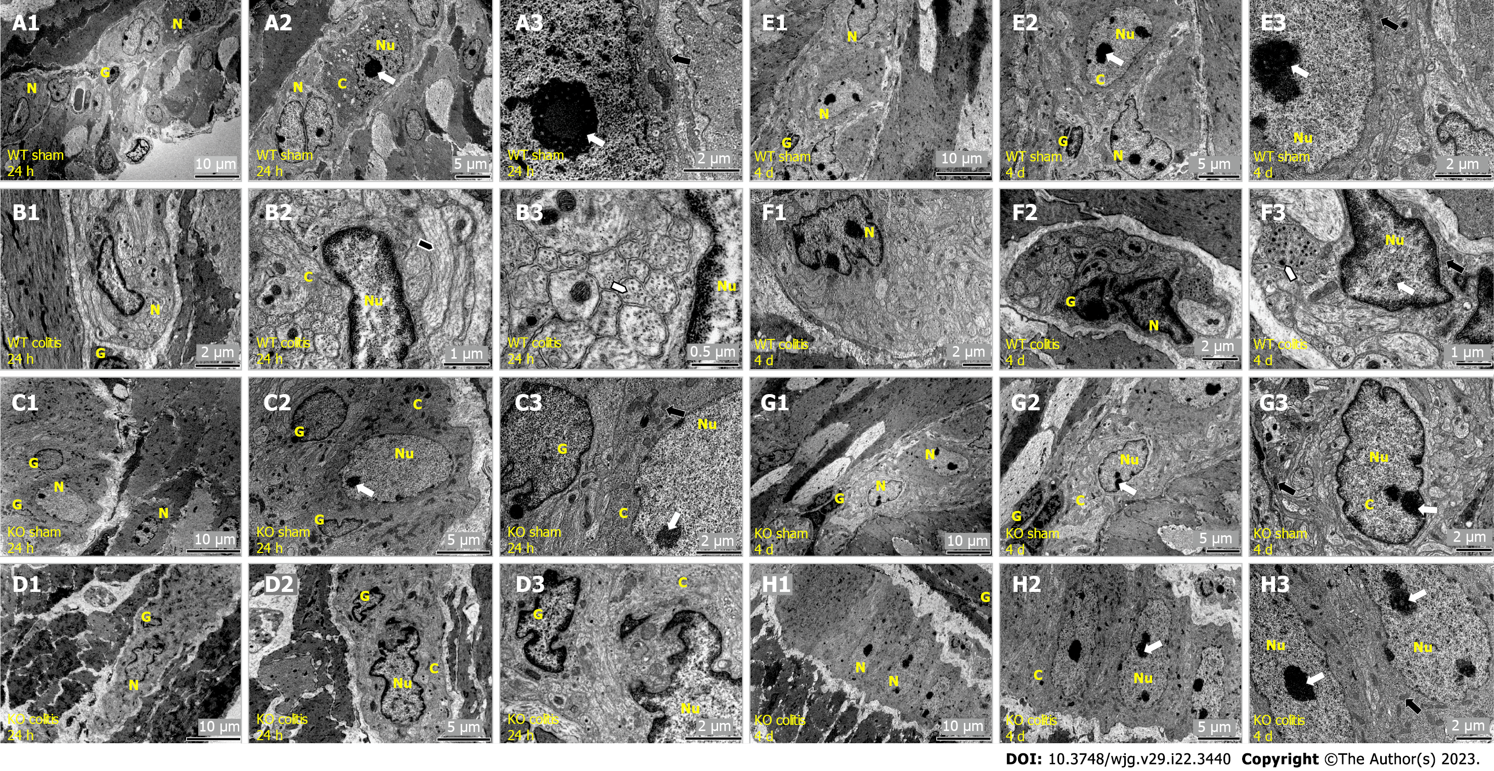
Figure 11 Transmission electron microscopy photomicrograph showing the ultrastructural organization of the myenteric plexus of the distal colon in mice.
A1-A3: Wild-type (WT) sham 24 h; B1-B3: WT colitis 24 h; C: Knockout (KO) sham 24 h; D1-D3: KO colitis 24 h; E1-E3: WT sham 4 d; F1-F3: WT colitis 4 d; G1-G3: KO sham 4 d; H1-H3: KO colitis 4 d groups. In the WT colitis 24 h and KO colitis 24 h groups, neurons with a necrotic phenotype characterized by nuclear disruption and apparent nuclear autolysis, chromatin fragmentation and condensation, and cytoplasmic disorganization were observed. Enteric glial cells also showed morphological changes. In the WT colitis 4 d group and less frequently in the KO colitis 4 d group, neurons showed irregular nuclei and nuclear membrane invaginations. The cell membrane remained intact, and apoptotic bodies were sometimes observed. Enteric glial cells also showed features consistent with cell apoptosis. In the WT sham 24 h, KO sham 24 h, WT sham 4 d and KO sham 4 d groups, neurons exhibited large and rounded/oval nuclei, and there were no signs of structural changes in the cytoplasm. N: Myenteric neuron; G: Enteric glial cells; C: Myenteric neuron cytoplasm; Nu: Myenteric neuron nucleus. Nucleolus (white arrow), mitochondria (black arrow), nuclear membrane invaginations (black arrowhead), and vesicles and microtubules (white arrowhead). WT: Wild-type; KO: Knockout.
- Citation: Magalhães HIR, Machado FA, Souza RF, Caetano MAF, Figliuolo VR, Coutinho-Silva R, Castelucci P. Study of the roles of caspase-3 and nuclear factor kappa B in myenteric neurons in a P2X7 receptor knockout mouse model of ulcerative colitis. World J Gastroenterol 2023; 29(22): 3440-3468
- URL: https://www.wjgnet.com/1007-9327/full/v29/i22/3440.htm
- DOI: https://dx.doi.org/10.3748/wjg.v29.i22.3440


