Copyright
©The Author(s) 2023.
World J Gastroenterol. Jun 14, 2023; 29(22): 3440-3468
Published online Jun 14, 2023. doi: 10.3748/wjg.v29.i22.3440
Published online Jun 14, 2023. doi: 10.3748/wjg.v29.i22.3440
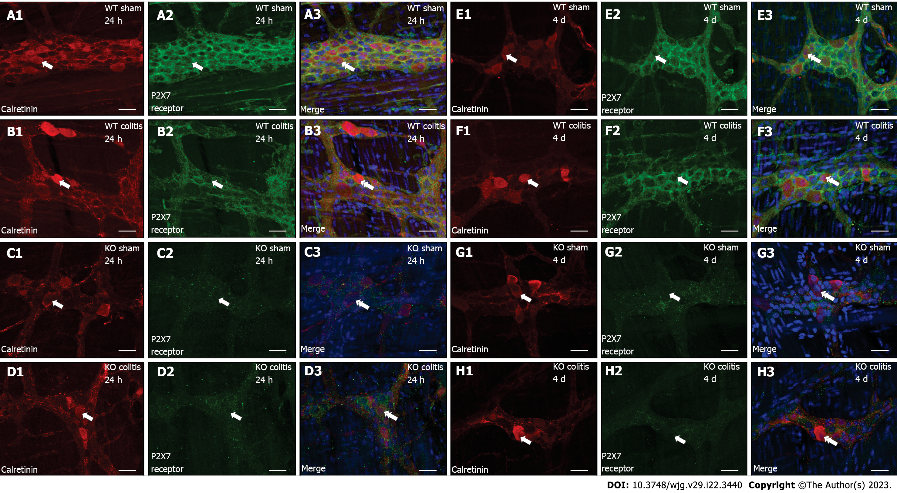
Figure 2 Colocalization of calretinin (red) with the P2X7 receptor (green) and DAPI (blue) in neurons in the myenteric plexus of the distal colon in mice.
A1-A3: Wild-type (WT) sham 24 h; B1-B3: WT colitis 24 h; C1-C3: Knockout (KO) sham 24 h; D1-D3: KO colitis 24 h; E1-E3: WT sham 4 d; F1-F3: WT colitis 4 d; G1-G3: KO sham 4 d; H1-H3: KO colitis 4 d groups. The single arrows indicate calretinin-ir (A1-H1) and P2X7 receptor-ir (A2-H2) neurons, and the double arrows indicate the colocalization of calretinin with the P2X7 receptor and DAPI (A3-H3). Scale bar = 30 µm. WT: Wild-type; KO: Knockout.
- Citation: Magalhães HIR, Machado FA, Souza RF, Caetano MAF, Figliuolo VR, Coutinho-Silva R, Castelucci P. Study of the roles of caspase-3 and nuclear factor kappa B in myenteric neurons in a P2X7 receptor knockout mouse model of ulcerative colitis. World J Gastroenterol 2023; 29(22): 3440-3468
- URL: https://www.wjgnet.com/1007-9327/full/v29/i22/3440.htm
- DOI: https://dx.doi.org/10.3748/wjg.v29.i22.3440


