Copyright
©The Author(s) 2023.
World J Gastroenterol. Jun 7, 2023; 29(21): 3328-3340
Published online Jun 7, 2023. doi: 10.3748/wjg.v29.i21.3328
Published online Jun 7, 2023. doi: 10.3748/wjg.v29.i21.3328
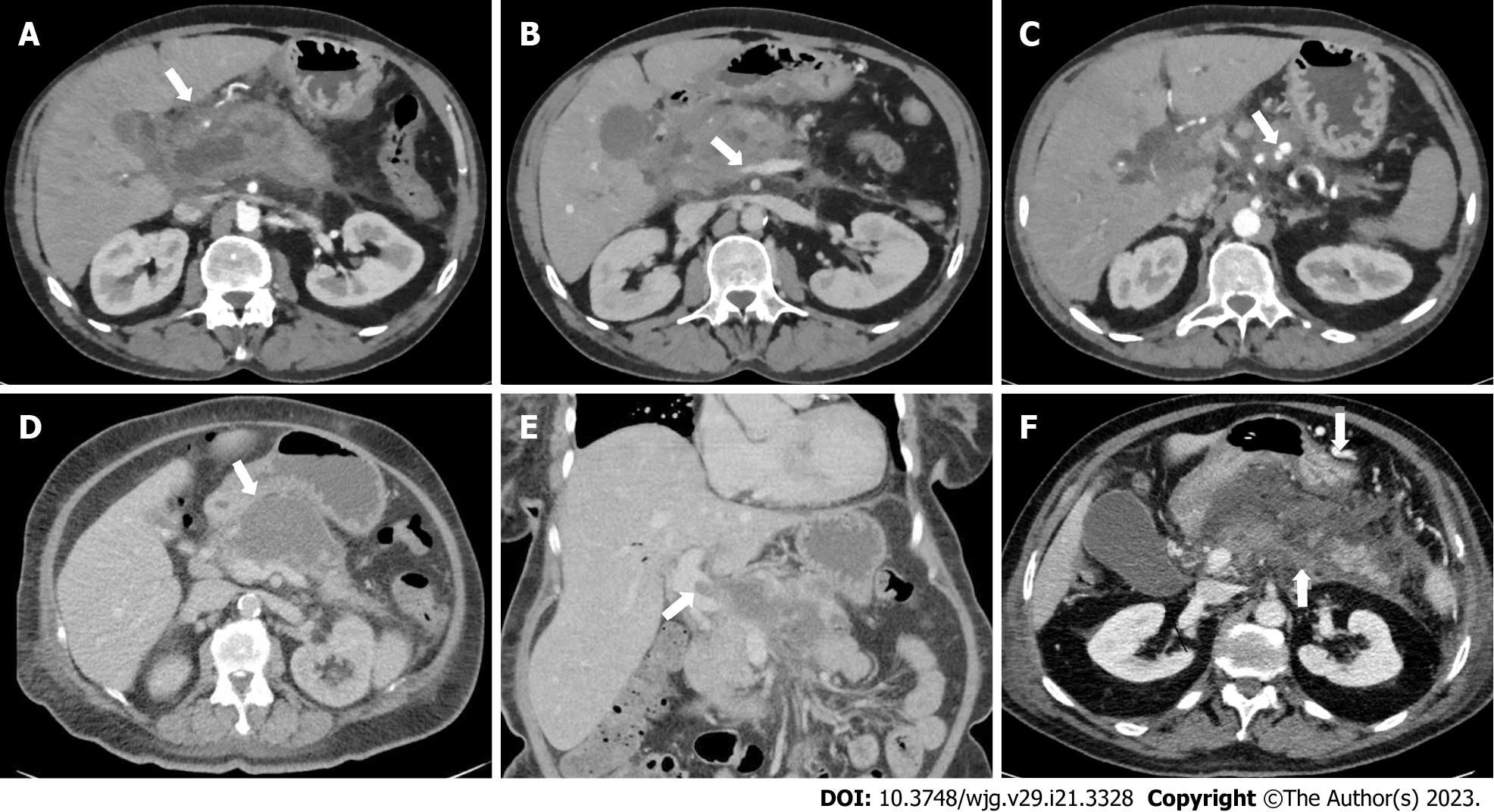
Figure 1 Imaging findings of case vignette.
A: Acute necrotic collection in the head of the pancreas in case vignette 1; B: Luminal narrowing of the portal vein without the presence of collateral circulation in case vignette 1; C: Pseudoaneurysm in the proximal splenic artery in case vignette 1; D: Almost fully encapsulated pancreatic necrosis without gas configurations in case vignette 2; E: Luminal filling defect in the portal vein without the presence of collateral circulation in case vignette 2; F: Extension of the thrombus to the splenic vein (arrow pointing upwards) and expansion of the collateral pathway in the gastroepiploic veins along the great curvature of the stomach (arrow pointing downwards) in case vignette 3.
- Citation: Sissingh NJ, Groen JV, Timmerhuis HC, Besselink MG, Boekestijn B, Bollen TL, Bonsing BA, Klok FA, van Santvoort HC, Verdonk RC, van Eijck CHJ, van Hooft JE, Mieog JSD. Therapeutic anticoagulation for splanchnic vein thrombosis in acute pancreatitis: A national survey and case-vignette study. World J Gastroenterol 2023; 29(21): 3328-3340
- URL: https://www.wjgnet.com/1007-9327/full/v29/i21/3328.htm
- DOI: https://dx.doi.org/10.3748/wjg.v29.i21.3328


