Copyright
©The Author(s) 2023.
World J Gastroenterol. May 28, 2023; 29(20): 3145-3156
Published online May 28, 2023. doi: 10.3748/wjg.v29.i20.3145
Published online May 28, 2023. doi: 10.3748/wjg.v29.i20.3145
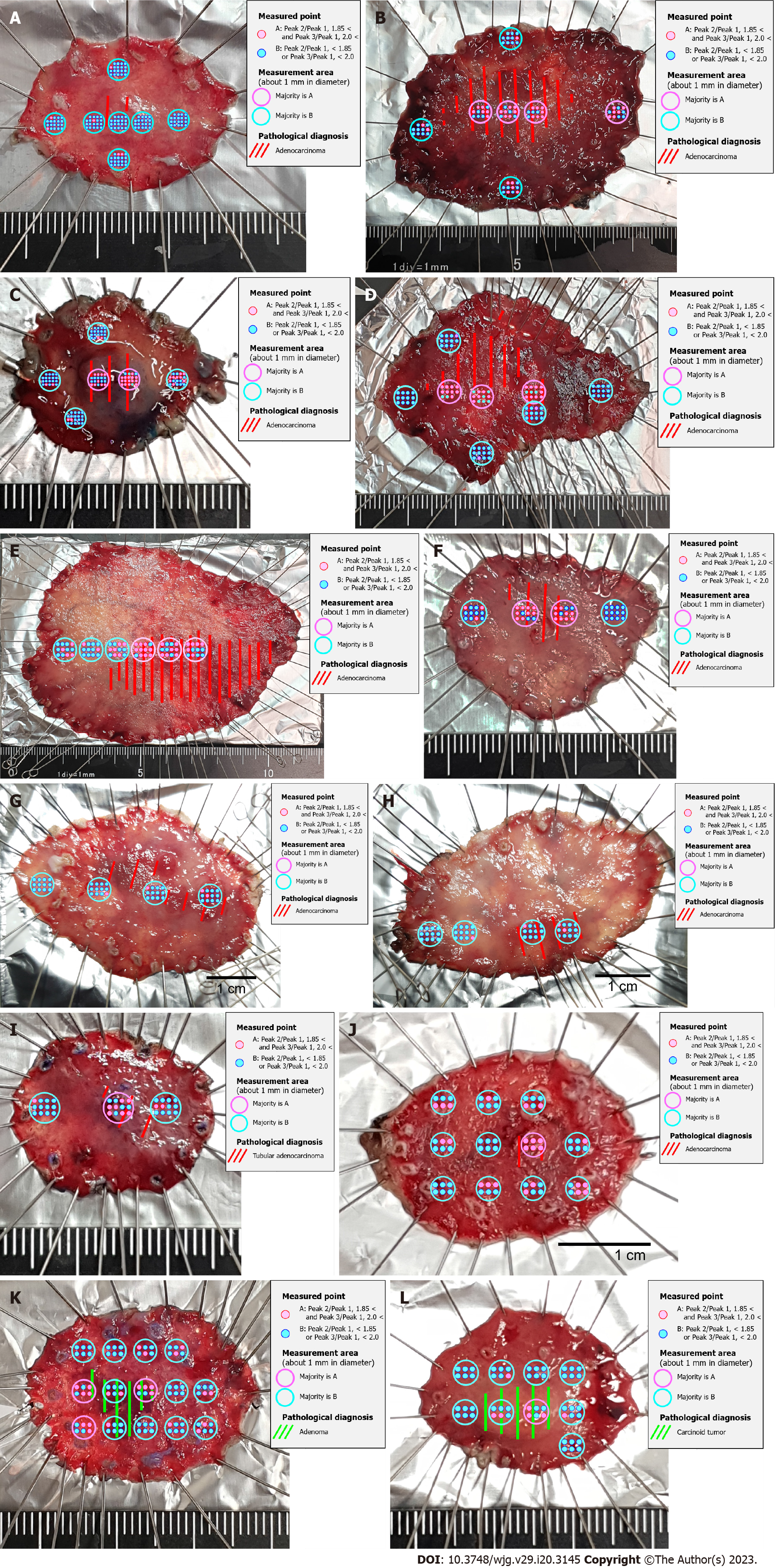
Figure 3 Comparison of Raman spectroscopic analysis and pathological diagnosis of sample Sto (well-differentiated tubular adenocarcinoma, pT1a-pT1b1; tubular adenoma; carcinoid tumor, NET G1).
A: Sto-1; B: Sto-2; C: Sto-3; D: Sto-4; E: Sto-5; F: Sto-6; G: Sto-7; H: Sto-8; I: Sto-9; J: Sto-10; K: Sto-11; L: Sto-12.
- Citation: Ito H, Uragami N, Miyazaki T, Shimamura Y, Ikeda H, Nishikawa Y, Onimaru M, Matsuo K, Isozaki M, Yang W, Issha K, Kimura S, Kawamura M, Yokoyama N, Kushima M, Inoue H. Determination of esophageal squamous cell carcinoma and gastric adenocarcinoma on raw tissue using Raman spectroscopy. World J Gastroenterol 2023; 29(20): 3145-3156
- URL: https://www.wjgnet.com/1007-9327/full/v29/i20/3145.htm
- DOI: https://dx.doi.org/10.3748/wjg.v29.i20.3145


