Copyright
©The Author(s) 2023.
World J Gastroenterol. May 28, 2023; 29(20): 3084-3102
Published online May 28, 2023. doi: 10.3748/wjg.v29.i20.3084
Published online May 28, 2023. doi: 10.3748/wjg.v29.i20.3084
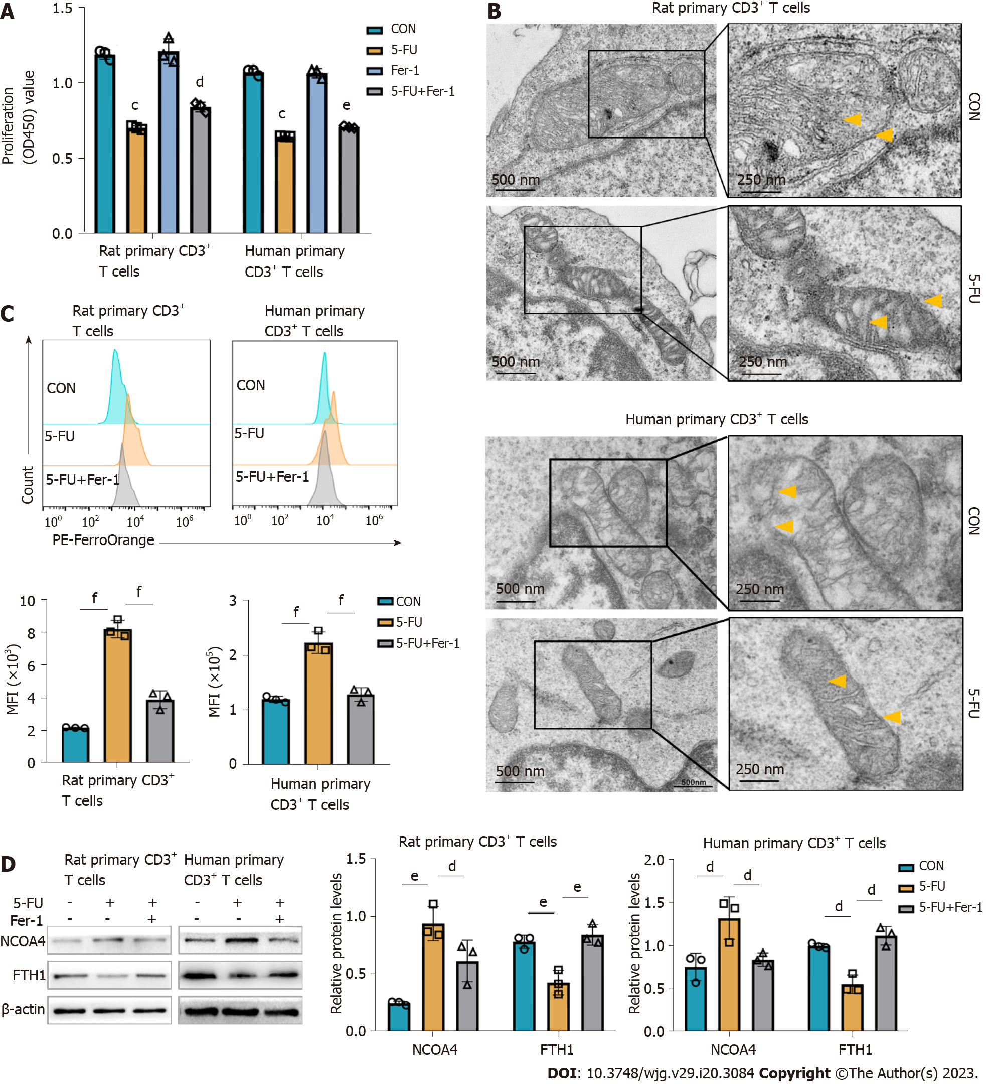
Figure 6 5-fluorouracil increases the pool of labile iron and contributes to ferroptosis in CD3+ T cells.
Vitro experiments were conducted on rat splenic and human peripheral blood CD3+ T cells, which were sorted by immunomagnetic beads. According to predetermined IC50, the rat, and human primary CD3+ T cells were treated with 5-fluorouracil (5-FU) (15 μmol/L) and ferrostatin-1 (Fer-1) (2 μmol/L) for 48 h. A: Cell viability was assessed with a Cell Counting Kit-8 kit; the results are expressed as optical density read at 450 nm; B: Transmission electron microscopy images of mitochondria in T cells show increased membrane density (yellow arrows) and a shrunk morphology (Scale bar, 500 nm, and 250 nm); C: Representative FACS data showing FerroOrange staining as the measurement of the level of cytoplasmic labile iron in primary CD3+ T cells. The graph shows the statistical analysis of mean fluorescence intensity in the different groups; D: Levels of ferritinophagy-related proteins nuclear receptor coactivator 4 and ferritin heavy chain 1 were assessed by western blot. CON groups: T cells were treated with DMSO for 48 h; 5-FU groups: T cells were treated with 5-FU (15 μmol/L) for 48 h; 5-FU+ Fer-1 groups: T cells were treated with 5-FU (15 μmol/L) and Fer-1 (2 μmol/L) for 48 h. Statistical analysis was done by one-way ANOVA, n = 3. Data are shown as mean ± SD. Data are shown as mean ± SD. cP < 0.001 vs the control group, dP < 0.05 vs the 5-FU group, eP < 0.01 vs the 5-FU group, fP < 0.001 vs the 5-FU group. CON: Untreated control groups; 5-FU: 5-fluorouracil; NCOA4: Nuclear receptor coactivator 4; FTH1: Ferritin heavy chain 1; Fer-1: Ferrostatin-1.
- Citation: Wang H, Wang ZL, Zhang S, Kong DJ, Yang RN, Cao L, Wang JX, Yoshida S, Song ZL, Liu T, Fan SL, Ren JS, Li JH, Shen ZY, Zheng H. Metronomic capecitabine inhibits liver transplant rejection in rats by triggering recipients’ T cell ferroptosis. World J Gastroenterol 2023; 29(20): 3084-3102
- URL: https://www.wjgnet.com/1007-9327/full/v29/i20/3084.htm
- DOI: https://dx.doi.org/10.3748/wjg.v29.i20.3084


