Copyright
©The Author(s) 2023.
World J Gastroenterol. May 14, 2023; 29(18): 2733-2746
Published online May 14, 2023. doi: 10.3748/wjg.v29.i18.2733
Published online May 14, 2023. doi: 10.3748/wjg.v29.i18.2733
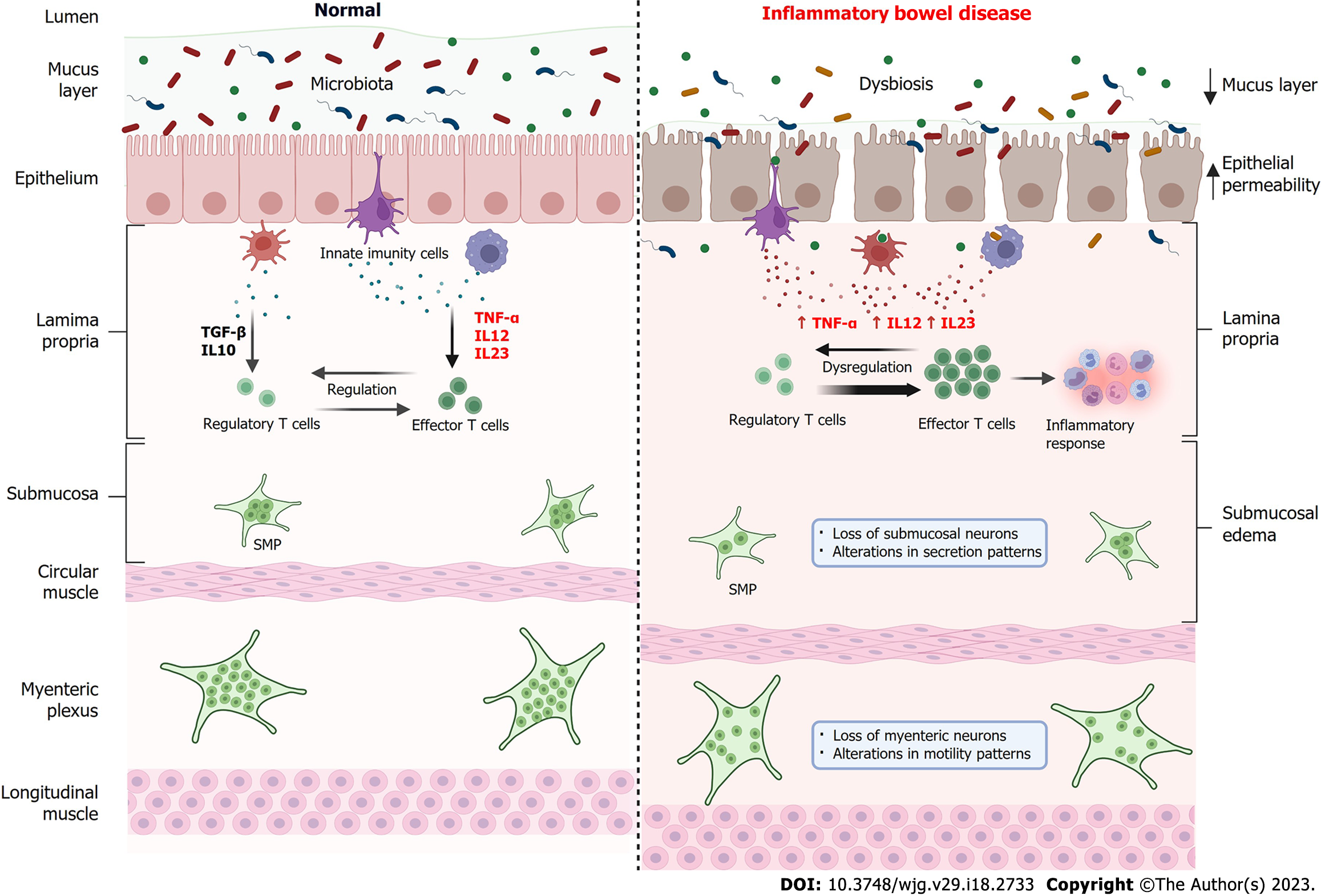
Figure 3 Comparison of the intestine in normal conditions and in the bowel with inflammatory bowel disease.
Under normal conditions (left), the intestinal microbiota establishes a symbiotic condition with the host, the intestinal barrier is intact and functional, and there is a balance between the levels of pro-inflammatory cytokines such as tumor necrosis factor alpha (TNF-α), interleukin (IL)-12 and IL-23 and anti-inflammatory cytokines such as transforming growth factor beta and IL-10 by innate immune cells. In this way, there is a regulation of the immune response through a balance between regulatory T cells and effector T cells. Submucosal plexus (SMP) and myenteric plexus neurons are functional and controlling, respectively, fluid secretion and intestinal motility. In inflammatory bowel diseases (IBD) (right), there is an imbalance in the intestinal microbiota, the intestinal barrier is compromised, with a reduction in the mucous layer, increased epithelial permeability and consequent passage of microorganisms to the lamina propria. Innate immune cells increase the secretion of pro-inflammatory cytokines such as TNF-α, IL-12 and IL-23, which leads to a dysregulation of the immune system mediated by T cells, increasing the activity of effector T cells which, in turn, recruit cells for the inflammatory response. In IBD, submucosal edema is observed, as well as a reduction in the number of neurons in the SMP, causing changes in secretion patterns and loss of neurons in the myenteric plexus, resulting in changes in motility patterns. TNF-α: Tumor necrosis factor-alpha; IL: Interleukin. Created with BioRender.com.
- Citation: Souza RF, Caetano MAF, Magalhães HIR, Castelucci P. Study of tumor necrosis factor receptor in the inflammatory bowel disease. World J Gastroenterol 2023; 29(18): 2733-2746
- URL: https://www.wjgnet.com/1007-9327/full/v29/i18/2733.htm
- DOI: https://dx.doi.org/10.3748/wjg.v29.i18.2733


