Copyright
©The Author(s) 2023.
World J Gastroenterol. May 7, 2023; 29(17): 2642-2656
Published online May 7, 2023. doi: 10.3748/wjg.v29.i17.2642
Published online May 7, 2023. doi: 10.3748/wjg.v29.i17.2642
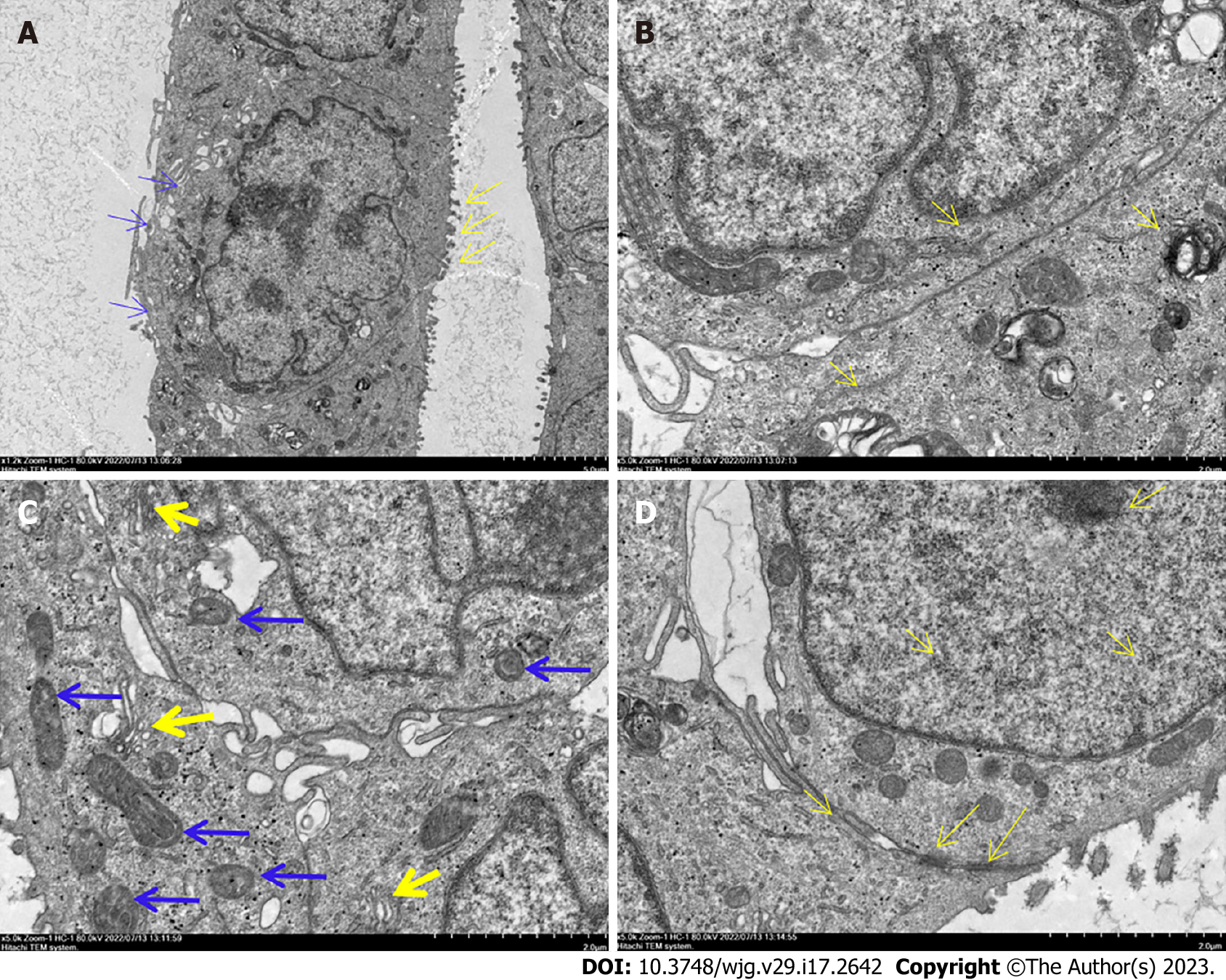
Figure 3 Ultrastructure of DPC-X1 under a transmission electron microscope.
A: DPC-X1 has a large and deformed nucleus; it is increased in number. The nucleolus is clustered in the nuclear membrane. There are fewer cytoplasm. Microvilli (yellow arrow) and pseudopodia (blue arrow) were visible on the cell surface; B: DPC-X1 cells are rich in the endoplasmic reticulum (yellow arrow) and ribosome; C: DPC-X1 cells have well-developed Golgi apparatus (yellow arrow), and the size and shape of the mitochondria are different (blue arrow); D: DPC-X1 desmosome structure can be seen between the cells (arrow).
- Citation: Xu H, Chai CP, Miao X, Tang H, Hu JJ, Zhang H, Zhou WC. Establishment and characterization of a new human ampullary carcinoma cell line, DPC-X1. World J Gastroenterol 2023; 29(17): 2642-2656
- URL: https://www.wjgnet.com/1007-9327/full/v29/i17/2642.htm
- DOI: https://dx.doi.org/10.3748/wjg.v29.i17.2642


