Copyright
©The Author(s) 2023.
World J Gastroenterol. May 7, 2023; 29(17): 2600-2615
Published online May 7, 2023. doi: 10.3748/wjg.v29.i17.2600
Published online May 7, 2023. doi: 10.3748/wjg.v29.i17.2600
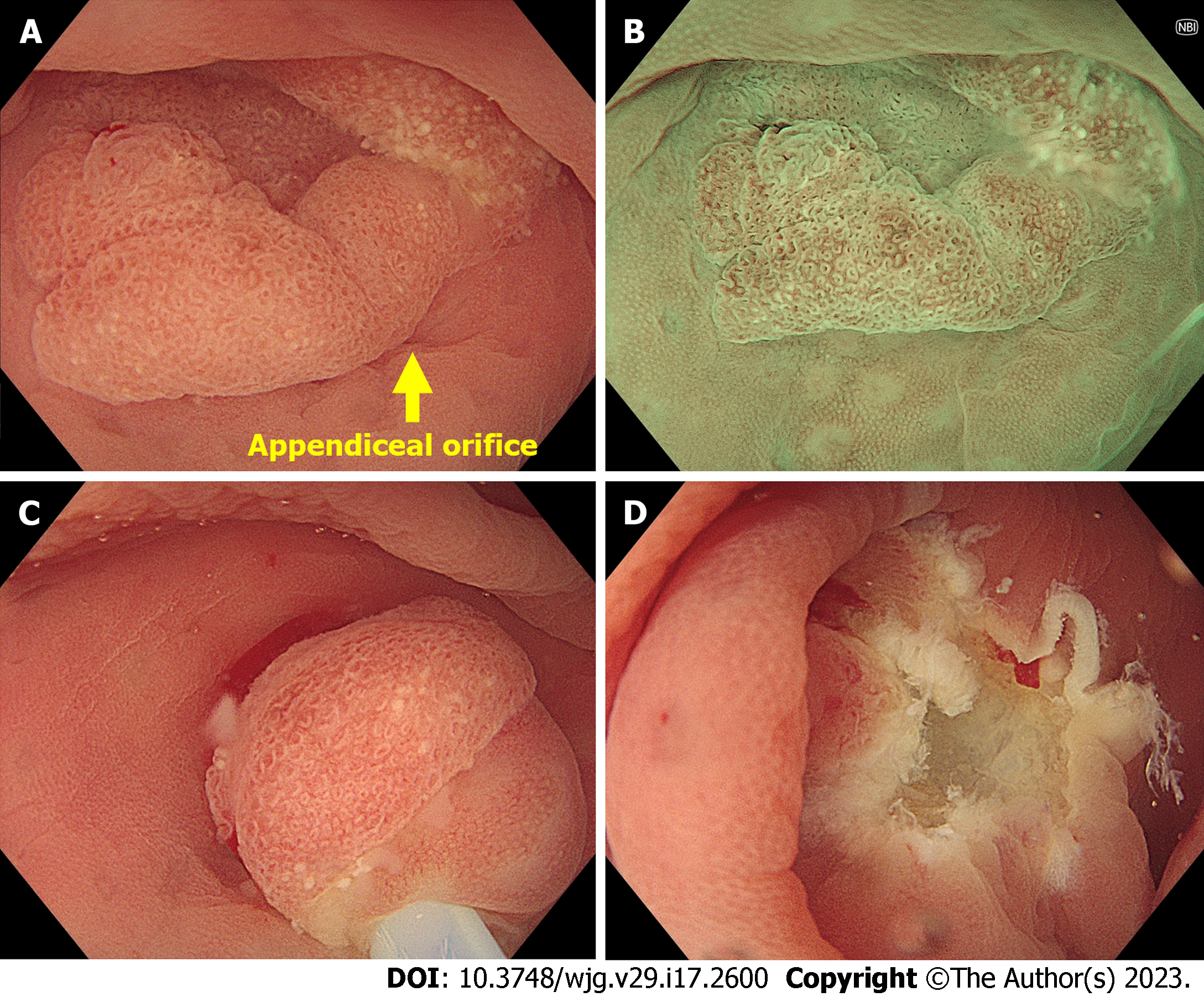
Figure 5 Underwater endoscopic mucosal resection for a polyp involving appendiceal orifice.
A: Endoscopic image showing a 15-mm sessile polyp involving the appendiceal orifice; B: Magnifying narrow-band image showing a type 1 polyp of the Japan Narrow-band Imaging Expert Team classification with open pit pattern. The most likely diagnosis was sessile serrated lesion; C: Underwater snaring without submucosal injection; D: Mucosal defect after completed underwater endoscopic mucosal resection.
- Citation: Pattarajierapan S, Takamaru H, Khomvilai S. Difficult colorectal polypectomy: Technical tips and recent advances. World J Gastroenterol 2023; 29(17): 2600-2615
- URL: https://www.wjgnet.com/1007-9327/full/v29/i17/2600.htm
- DOI: https://dx.doi.org/10.3748/wjg.v29.i17.2600


