Copyright
©The Author(s) 2023.
World J Gastroenterol. Mar 28, 2023; 29(12): 1875-1898
Published online Mar 28, 2023. doi: 10.3748/wjg.v29.i12.1875
Published online Mar 28, 2023. doi: 10.3748/wjg.v29.i12.1875
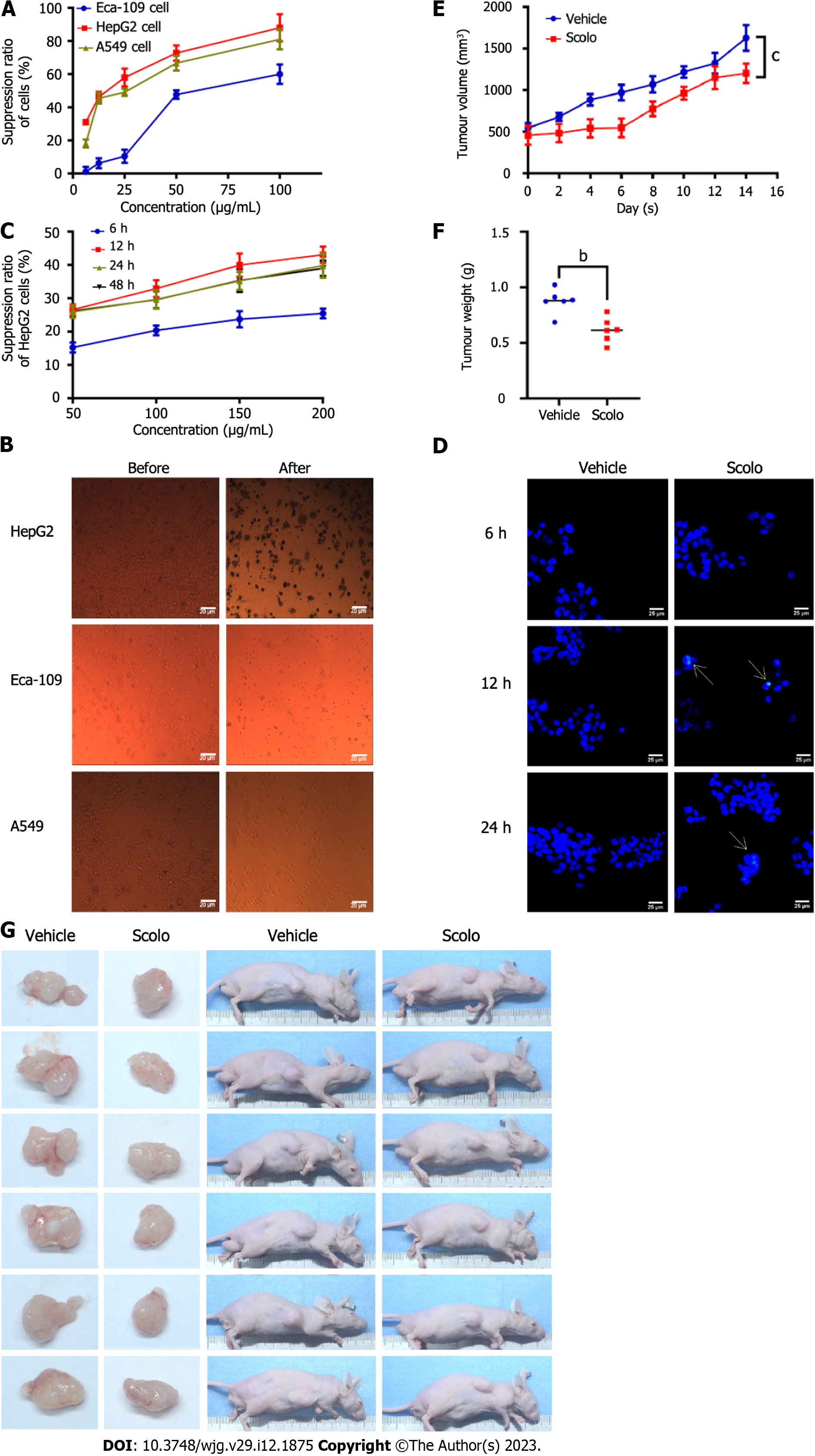
Figure 3 Antihepatoma effect of scolopentide.
A: The CCK8 assay showed the suppression ratio of Eca-109, HepG2, and A549 cells treated with extracted scolopentide at different concentrations; B: Morphological changes in Eca-109, HepG2, and A549 cells under a light microscope (× 40); after treatment with extracted scolopentide, three cells were morphologically changed, especially HepG2 cells; C: The CCK8 assay showed the suppression ratio of HepG2 cells treated with synthetic scolopentide at different times (6 h, 12 h, 24 h, and 48 h) and different concentrations (50 μg/mL, 100 μg/mL, 150 μg/mL, and 200 μg/mL); D: Hoechst 33342 staining (× 400) of HepG2 cells. After treatment with synthetic scolopentide for 12 h and 24 h, cytoplasmic highlight staining and nuclear pyknosis occurred; E-G: Tumor volume and weight of the scolopentide group (synthetic scolopentide 500 mg/kg/d) and vehicle group (constant volume of normal saline). n = 6, bP < 0.01, cP < 0.001.
- Citation: Hu YX, Liu Z, Zhang Z, Deng Z, Huang Z, Feng T, Zhou QH, Mei S, Yi C, Zhou Q, Zeng PH, Pei G, Tian S, Tian XF. Antihepatoma peptide, scolopentide, derived from the centipede scolopendra subspinipes mutilans. World J Gastroenterol 2023; 29(12): 1875-1898
- URL: https://www.wjgnet.com/1007-9327/full/v29/i12/1875.htm
- DOI: https://dx.doi.org/10.3748/wjg.v29.i12.1875


