Copyright
©The Author(s) 2023.
World J Gastroenterol. Mar 28, 2023; 29(12): 1852-1862
Published online Mar 28, 2023. doi: 10.3748/wjg.v29.i12.1852
Published online Mar 28, 2023. doi: 10.3748/wjg.v29.i12.1852
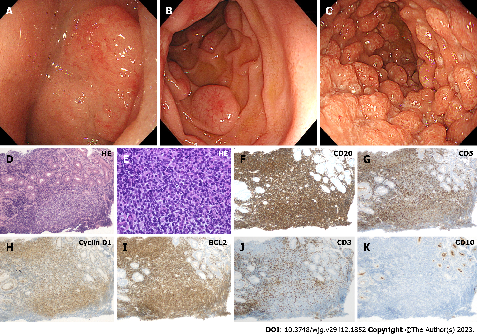
Figure 6 Representative endoscopic and pathological images of duodenal mantle cell lymphoma (Cases 10–12).
A: Case 10. Elevated lesion with erosions on the surface in the duodenum; B: Case 11. Typical morphology of mantle cell lymphoma showing multiple lymphomatous polyposis in the duodenum. Subepithelial lesion-like protruded lesions accompanying dilated vasculature on the surface are seen; C: Case 12. Numerous, diffuse polypoid lesions in the duodenum; D–K: Pathological images of Case 12. Lymphoma cells retain a homogeneous pattern of cell size on hematoxylin and eosin stain (D: × 10, E: × 40). Cells are positive for CD20, CD5, Cyclin D1, BCL2, while negative for CD3 and CD10. HE: Hematoxylin and eosin.
- Citation: Iwamuro M, Tanaka T, Okada H. Review of lymphoma in the duodenum: An update of diagnosis and management. World J Gastroenterol 2023; 29(12): 1852-1862
- URL: https://www.wjgnet.com/1007-9327/full/v29/i12/1852.htm
- DOI: https://dx.doi.org/10.3748/wjg.v29.i12.1852


