Copyright
©The Author(s) 2023.
World J Gastroenterol. Mar 28, 2023; 29(12): 1852-1862
Published online Mar 28, 2023. doi: 10.3748/wjg.v29.i12.1852
Published online Mar 28, 2023. doi: 10.3748/wjg.v29.i12.1852
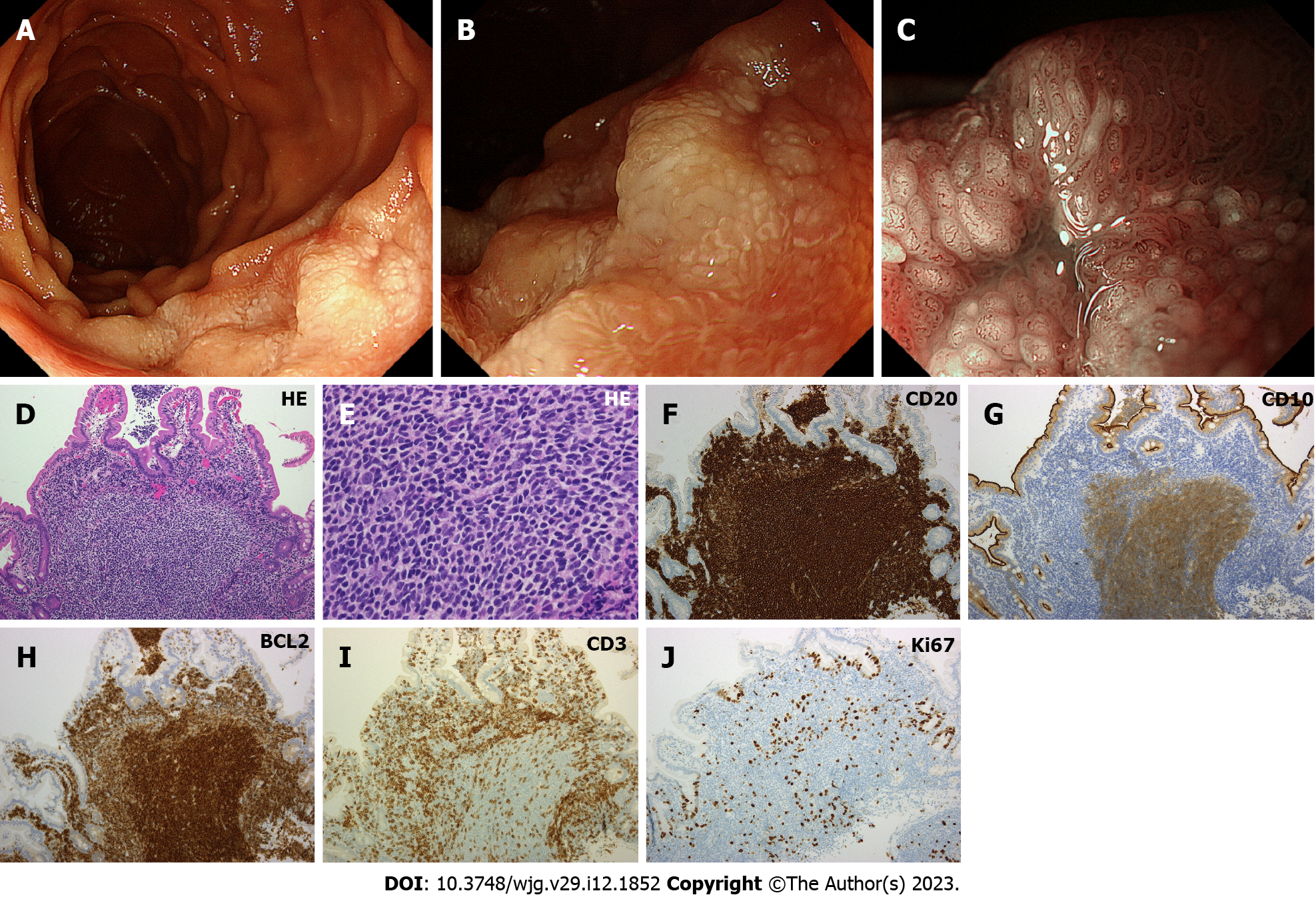
Figure 3 Representative endoscopic and pathological images of duodenal follicular lymphoma (Case 3).
A: Esophagogastroduodenoscopy shows thick, white mucosa in the descending portion of the duodenum; B: Magnifying observation reveals enlarged whitish villi; C: Magnifying observation with narrow band imaging emphasizes enlarged villi with elongated vasculature on the surface; D–J: Pathological images of the biopsied specimen. Hematoxylin and eosin stain shows medium-sized tumor cells that form follicular structures and diffusely invade into the villi (D: × 10, E: × 40). Lymphoma cells are positive for CD20, CD10, and BCL2, while negative for CD3. Tumor cells are sparsely positive for Ki67. HE: Hematoxylin and eosin.
- Citation: Iwamuro M, Tanaka T, Okada H. Review of lymphoma in the duodenum: An update of diagnosis and management. World J Gastroenterol 2023; 29(12): 1852-1862
- URL: https://www.wjgnet.com/1007-9327/full/v29/i12/1852.htm
- DOI: https://dx.doi.org/10.3748/wjg.v29.i12.1852


