Copyright
©The Author(s) 2022.
World J Gastroenterol. Feb 14, 2022; 28(6): 675-682
Published online Feb 14, 2022. doi: 10.3748/wjg.v28.i6.675
Published online Feb 14, 2022. doi: 10.3748/wjg.v28.i6.675
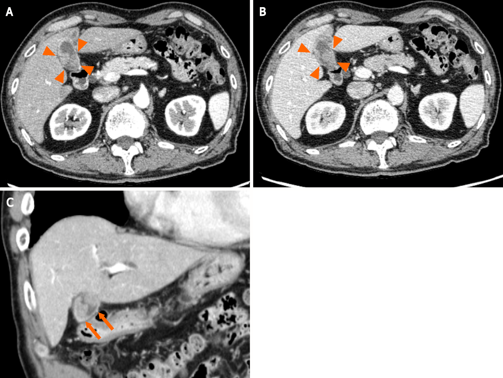
Figure 1 A contrast-enhanced abdominal computed tomography scan shows two irregular and highly contrast-enhanced masses (arrowheads and arrow) at the neck and body of the gallbladder as well as periportal lymph node enlargement, which is consistent with gallbladder cancer lymph node metastasis.
A: Axial section image in the early phase showing neck of the gallbladder; B: Axial section image in the delayed phase showing neck of the gallbladder in the delayed phase; C: Coronal sectional image showing body of the gallbladder.
- Citation: Hosoda K, Shimizu A, Kubota K, Notake T, Hayashi H, Yasukawa K, Umemura K, Kamachi A, Goto T, Tomida H, Yamazaki S, Narusawa Y, Asano N, Uehara T, Soejima Y. Gallbladder Burkitt’s lymphoma mimicking gallbladder cancer: A case report. World J Gastroenterol 2022; 28(6): 675-682
- URL: https://www.wjgnet.com/1007-9327/full/v28/i6/675.htm
- DOI: https://dx.doi.org/10.3748/wjg.v28.i6.675


