Copyright
©The Author(s) 2022.
World J Gastroenterol. Feb 7, 2022; 28(5): 594-601
Published online Feb 7, 2022. doi: 10.3748/wjg.v28.i5.594
Published online Feb 7, 2022. doi: 10.3748/wjg.v28.i5.594
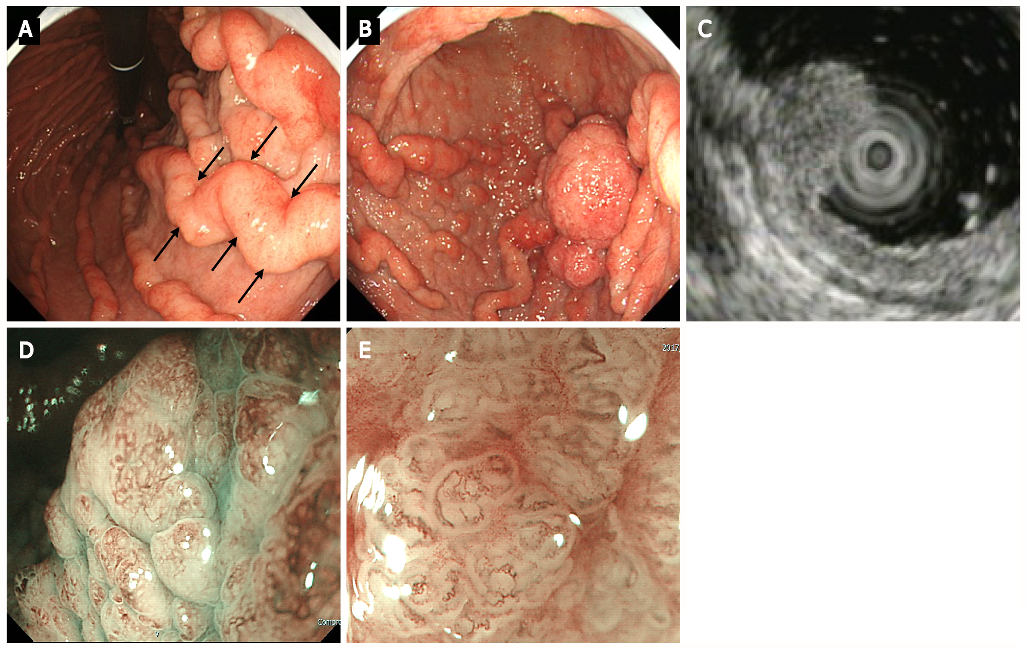
Figure 2 Upper gastrointestinal endoscopy.
A: Upper gastrointestinal endoscopy shows marked fold enlargement from the ventricular angle to the fundus (black arrow); B: A 40-mm broad-based protruding lesion is observed near the posterior wall of the greater curvature of the lower gastric body; C: Endoscopic ultrasonography shows the five layers of the gastric wall. In the lesion area, hypertrophy was observed in the first two layers. No noticeable structural changes were evident in the third layer or deeper; D: Low-magnification narrow-band imaging (NBI) shows granular surfaces with various sizes/forms and dilated vessels; E: High-magnification NBI demonstrates irregular microstructures with various forms and tortuous microvessels with changes in caliber.
- Citation: Fukushi K, Goda K, Kino H, Kondo M, Kanazawa M, Kashima K, Kanamori A, Abe K, Suzuki T, Tominaga K, Yamagishi H, Irisawa A. Curative resection with endoscopic submucosal dissection of early gastric cancer in Helicobacter pylori-negative Ménétrier’s disease: A case report. World J Gastroenterol 2022; 28(5): 594-601
- URL: https://www.wjgnet.com/1007-9327/full/v28/i5/594.htm
- DOI: https://dx.doi.org/10.3748/wjg.v28.i5.594


