Copyright
©The Author(s) 2022.
World J Gastroenterol. Feb 7, 2022; 28(5): 547-569
Published online Feb 7, 2022. doi: 10.3748/wjg.v28.i5.547
Published online Feb 7, 2022. doi: 10.3748/wjg.v28.i5.547
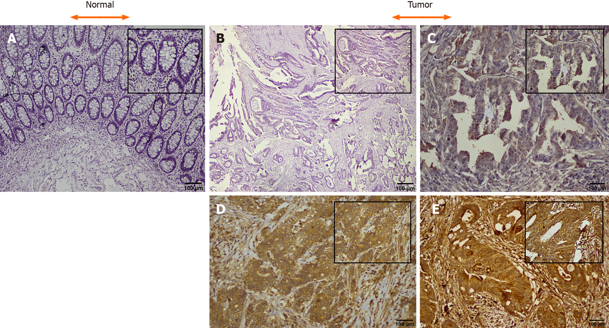
Figure 2 Representative images of immunohistochemical expression of the connective tissue growth factor protein in colorectal cancer and adjacent normal (H & E and DAB Chromogen × 100 insect × 400).
A: Negative control; B: Tumor negative staining; C: Tumor + 1, weak staining; D: Tumor + 2, moderate staining; E: Tumor + 3, strong staining. Scale bars: 100 μM.
- Citation: Bhat IP, Rather TB, Maqbool I, Rashid G, Akhtar K, Bhat GA, Parray FQ, Syed B, Khan IY, Kazi M, Hussain MD, Syed M. Connective tissue growth factor expression hints at aggressive nature of colorectal cancer. World J Gastroenterol 2022; 28(5): 547-569
- URL: https://www.wjgnet.com/1007-9327/full/v28/i5/547.htm
- DOI: https://dx.doi.org/10.3748/wjg.v28.i5.547


