Copyright
©The Author(s) 2022.
World J Gastroenterol. Dec 21, 2022; 28(47): 6769-6787
Published online Dec 21, 2022. doi: 10.3748/wjg.v28.i47.6769
Published online Dec 21, 2022. doi: 10.3748/wjg.v28.i47.6769
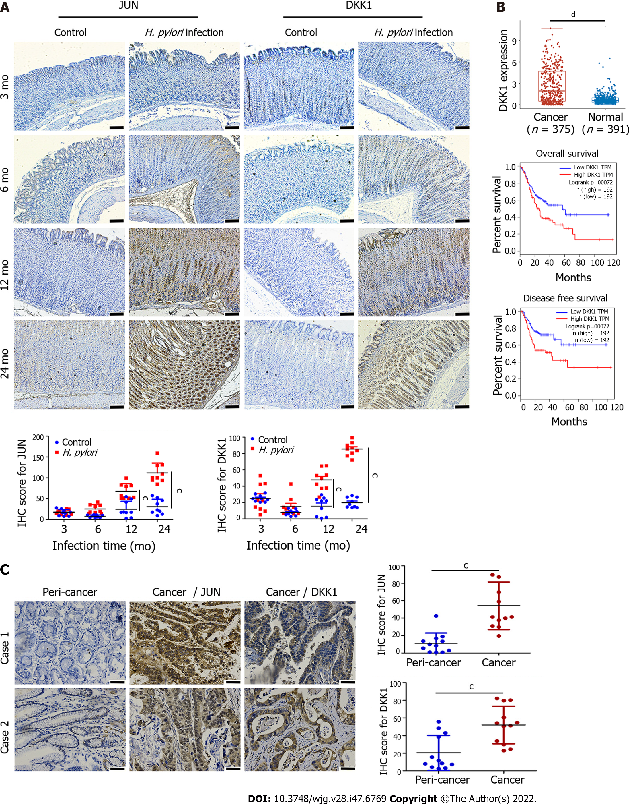
Figure 3 Immunohistochemistry for JUN and dickkopf-related protein 1 in gerbil stomach infected with Helicobacterpylori and gastric cancer tissues.
A: Immunohistochemical analysis of JUN and dickkopf-related protein 1 (DKK1) proteins in Helicobacter pylori (H. pylori)-infected gerbil stomach tissues at 3 mo, 6 mo, 12 mo, and 24 mo post-infection (n = 3). Dot diagrams show the quantification of JUN and DKK1 staining in immunohistochemical samples. Nine sections were chosen randomly from three different samples and used to determine mean ± SD of the immunohistochemistry (IHC) score, which was calculated as described in the methods. Scale bar = 100 μm. cP < 0.001; B: DKK1 expression and survival analysis in gastric cancer patients from the TCGA-STAD dataset. dP < 0.0001; C: Representative images of immunohistochemical staining of JUN and DKK1 proteins in human gastric cancer specimens. Dot diagrams show the quantitation of JUN and DKK1 staining. Scale bar = 50 μm. Data from 12 clinical samples of gastric cancer patients are expressed as mean ± SD. cP < 0.001. DKK1: Dickkopf-related protein 1; H. pylori: Helicobacter pylori.
- Citation: Luo M, Chen YJ, Xie Y, Wang QR, Xiang YN, Long NY, Yang WX, Zhao Y, Zhou JJ. Dickkopf-related protein 1/cytoskeleton-associated protein 4 signaling activation by Helicobacter pylori-induced activator protein-1 promotes gastric tumorigenesis via the PI3K/AKT/mTOR pathway. World J Gastroenterol 2022; 28(47): 6769-6787
- URL: https://www.wjgnet.com/1007-9327/full/v28/i47/6769.htm
- DOI: https://dx.doi.org/10.3748/wjg.v28.i47.6769


