Copyright
©The Author(s) 2022.
World J Gastroenterol. Dec 14, 2022; 28(46): 6564-6572
Published online Dec 14, 2022. doi: 10.3748/wjg.v28.i46.6564
Published online Dec 14, 2022. doi: 10.3748/wjg.v28.i46.6564
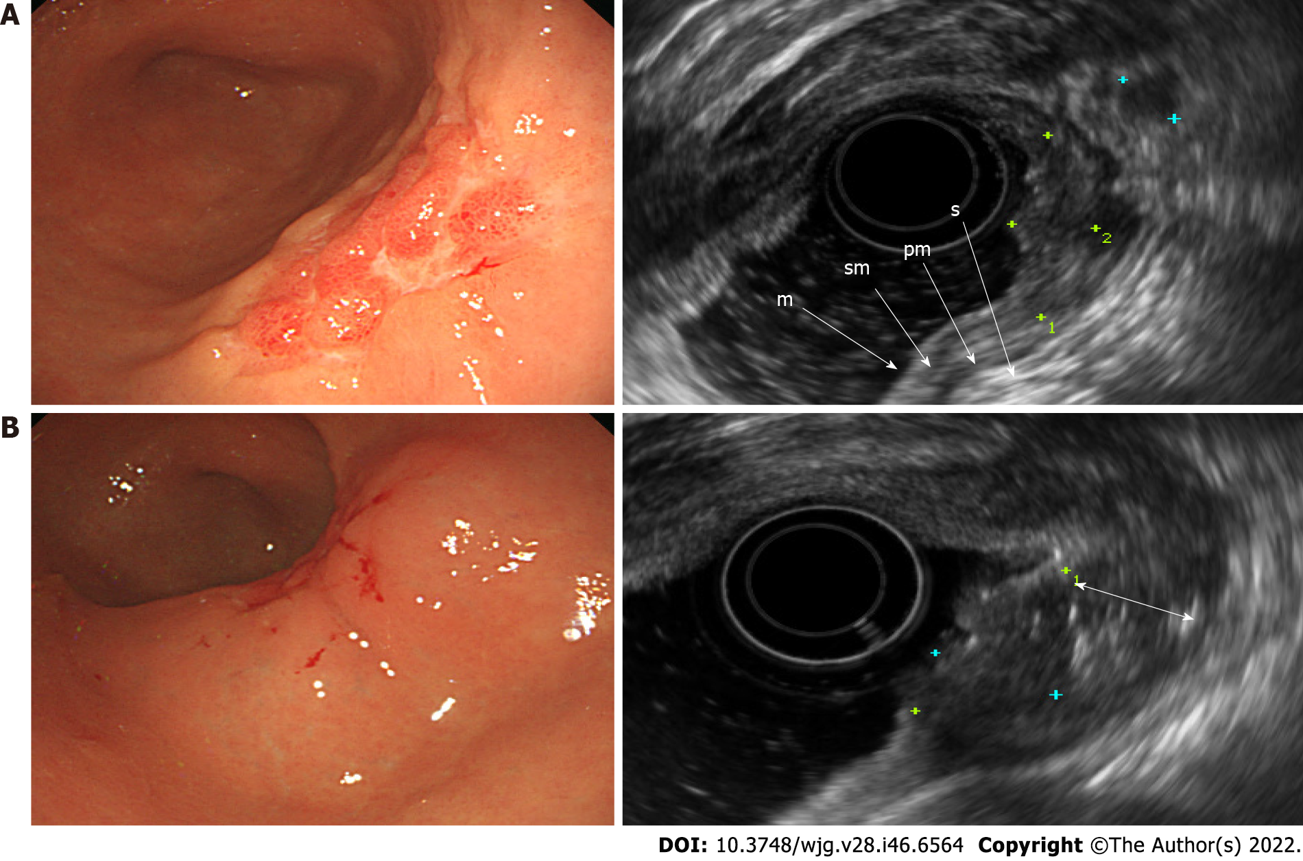
Figure 2 Endoscopic and ultrasonographic images and associated schematic diagrams of T1b early gastric cancer.
A: Standard endoscopic ultrasonography (EUS) showing that it is difficult to differentiate the extent of invasion from the mucosal layer to the submucosal layer; B: EUS after submucosal saline injection (SSI) showing clearly the boundary between the mucosa and the submucosa, meaning that the lesion, its infiltration depth into the mucosa, and the submucosa can be easily identified. m: Mucosa; sm: Submucosa; pm: Proper muscle; s: Serosa; double arrow, saline layer.
- Citation: Park JY, Jeon TJ. Diagnostic evaluation of endoscopic ultrasonography with submucosal saline injection for differentiating between T1a and T1b early gastric cancer. World J Gastroenterol 2022; 28(46): 6564-6572
- URL: https://www.wjgnet.com/1007-9327/full/v28/i46/6564.htm
- DOI: https://dx.doi.org/10.3748/wjg.v28.i46.6564


