Copyright
©The Author(s) 2022.
World J Gastroenterol. Dec 14, 2022; 28(46): 6564-6572
Published online Dec 14, 2022. doi: 10.3748/wjg.v28.i46.6564
Published online Dec 14, 2022. doi: 10.3748/wjg.v28.i46.6564
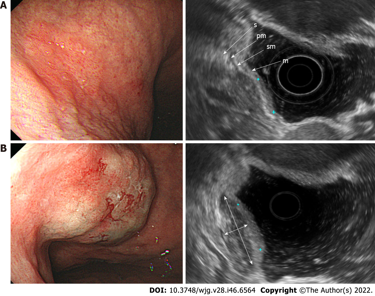
Figure 1 Endoscopic and ultrasonographic images and associated schematic diagrams of T1a early gastric cancer.
A: Standard endoscopic ultrasonography (EUS) showing that it is difficult to differentiate the extent of invasion from the mucosal layer to the submucosal layer; B: EUS after submucosal saline injection showing clearly the boundary between the mucosa and the submucosa, meaning that the T1a stage can be easily identified. m: Mucosa; sm: Submucosa; pm: Proper muscle; s: Serosa; double arrow, saline layer.
- Citation: Park JY, Jeon TJ. Diagnostic evaluation of endoscopic ultrasonography with submucosal saline injection for differentiating between T1a and T1b early gastric cancer. World J Gastroenterol 2022; 28(46): 6564-6572
- URL: https://www.wjgnet.com/1007-9327/full/v28/i46/6564.htm
- DOI: https://dx.doi.org/10.3748/wjg.v28.i46.6564


