Copyright
©The Author(s) 2022.
World J Gastroenterol. Dec 14, 2022; 28(46): 6512-6521
Published online Dec 14, 2022. doi: 10.3748/wjg.v28.i46.6512
Published online Dec 14, 2022. doi: 10.3748/wjg.v28.i46.6512
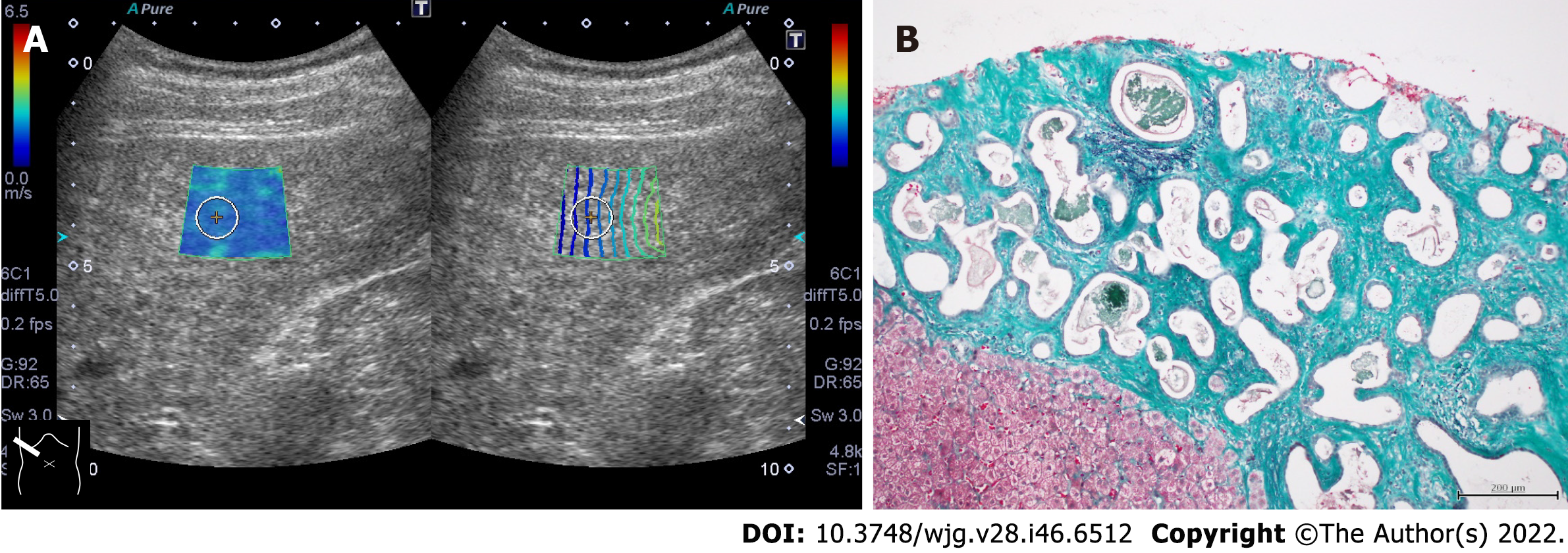
Figure 3 Representative case of von Meyenburg complex.
A: The liver showed a coarse parenchymal texture on ultrasound, and its shear wave propagation velocity was 1.86 m/s (10.3 kPa); B: Ultrasound-guided liver biopsy revealed bile duct proliferation with irregularly dilated small nuclei at the margins and fibrous stroma around the ducts. The fibrous stroma stained green on elastic Masson staining and was confirmed to be vitreous, confirming the diagnosis of von Meyenburg complex (ElasticaMasson staining, × 20).
- Citation: Naganuma H, Ishida H. Factors other than fibrosis that increase measured shear wave velocity. World J Gastroenterol 2022; 28(46): 6512-6521
- URL: https://www.wjgnet.com/1007-9327/full/v28/i46/6512.htm
- DOI: https://dx.doi.org/10.3748/wjg.v28.i46.6512


