Copyright
©The Author(s) 2022.
World J Gastroenterol. Dec 14, 2022; 28(46): 6512-6521
Published online Dec 14, 2022. doi: 10.3748/wjg.v28.i46.6512
Published online Dec 14, 2022. doi: 10.3748/wjg.v28.i46.6512
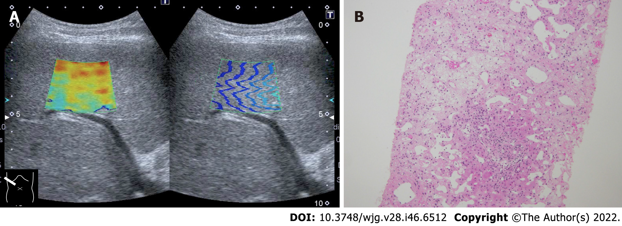
Figure 2 Representative case of massive hepatic necrosis.
A: Shear wave propagation velocity was 3.01 m/s (27.2 kPa); B: Biopsy specimen showed that hepatocytes in the central venous zone were completely lost due to necrosis and were replaced by reticular fibers. Hepatocytes remained in the portal vein area, which was one-half to one-third of the total, indicating sub extensive hepatic necrosis. Hematoxylin-eosin staining histologic finding (× 20).
- Citation: Naganuma H, Ishida H. Factors other than fibrosis that increase measured shear wave velocity. World J Gastroenterol 2022; 28(46): 6512-6521
- URL: https://www.wjgnet.com/1007-9327/full/v28/i46/6512.htm
- DOI: https://dx.doi.org/10.3748/wjg.v28.i46.6512


