Copyright
©The Author(s) 2022.
World J Gastroenterol. Nov 7, 2022; 28(41): 5993-6001
Published online Nov 7, 2022. doi: 10.3748/wjg.v28.i41.5993
Published online Nov 7, 2022. doi: 10.3748/wjg.v28.i41.5993
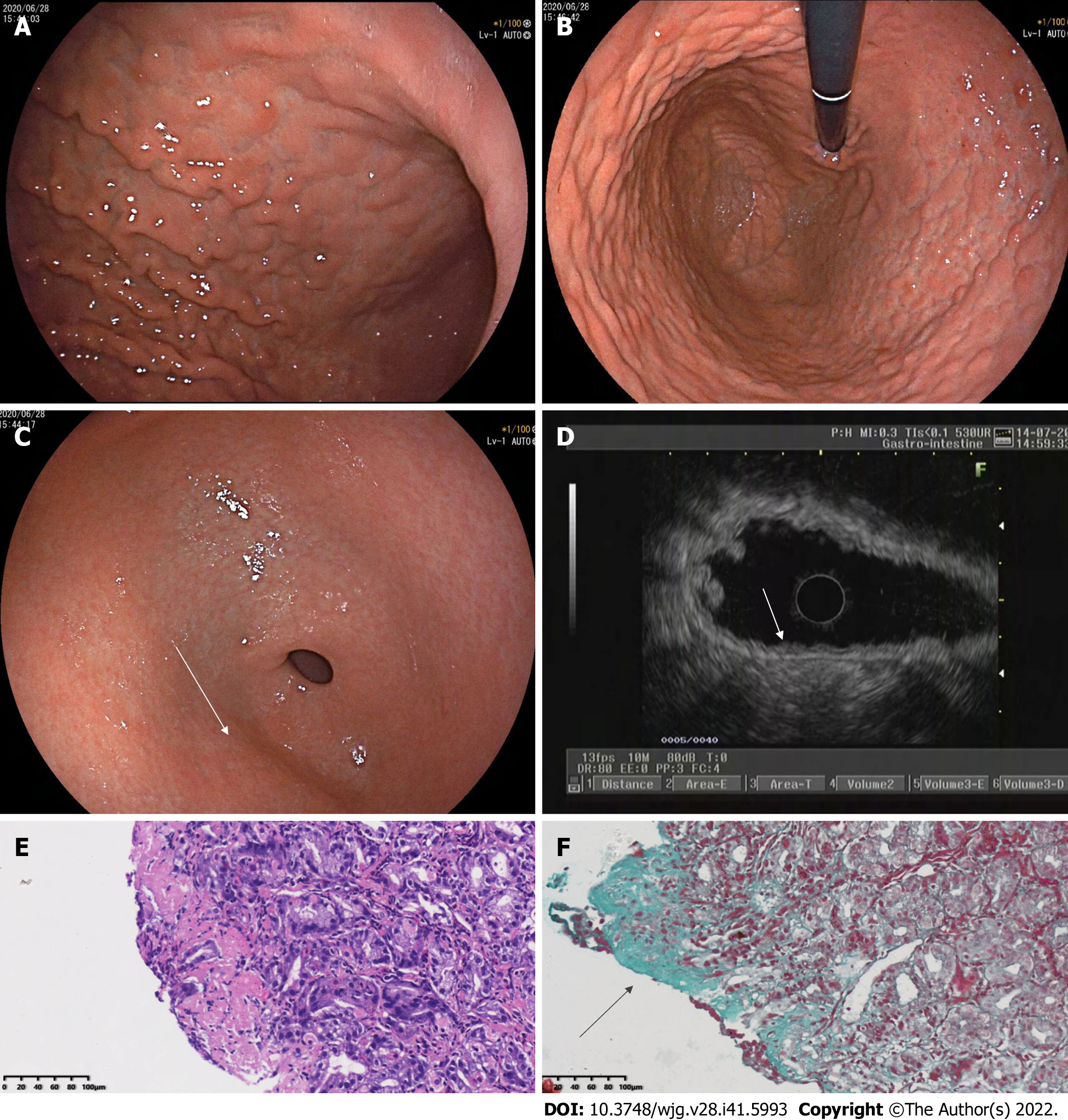
Figure 1 Findings of white light endoscopy, endoscopic ultrasound, and histopathology in 2020.
A: Esophagogastroduodenoscopy (EGD) in 2020 in a forward endoscopic view showed diffuse nodular elevation-depression changes in the mucosa of the entire gastric corpus; B: EGD in 2020 (in a retroflexed endoscopic view) showed diffuse nodular elevation-depression changes in the mucosa of the entire gastric corpus; C: EGD in 2020 showed nodular reddening-like changes in the anterior wall of the gastric antrum; D: Endoscopic ultrasound in 2020 showed an intact five-layer structure of the gastric corpus wall with hypoechoic changes in the mucosal layer, which is different from the echoes of normal mucosal layer; E: A biopsy of the gastric corpus in 2020 reported moderate chronic inflammation with erosion and mild activity; F: Masson staining of a gastric corpus specimen in 2020 showed collagen bands (blue areas).
- Citation: Zheng QH, Hu J, Yi XY, Xiao XH, Zhou LN, Li B, Bo XT. Collagenous gastritis in a young Chinese woman: A case report. World J Gastroenterol 2022; 28(41): 5993-6001
- URL: https://www.wjgnet.com/1007-9327/full/v28/i41/5993.htm
- DOI: https://dx.doi.org/10.3748/wjg.v28.i41.5993


