Copyright
©The Author(s) 2022.
World J Gastroenterol. Nov 7, 2022; 28(41): 5893-5909
Published online Nov 7, 2022. doi: 10.3748/wjg.v28.i41.5893
Published online Nov 7, 2022. doi: 10.3748/wjg.v28.i41.5893
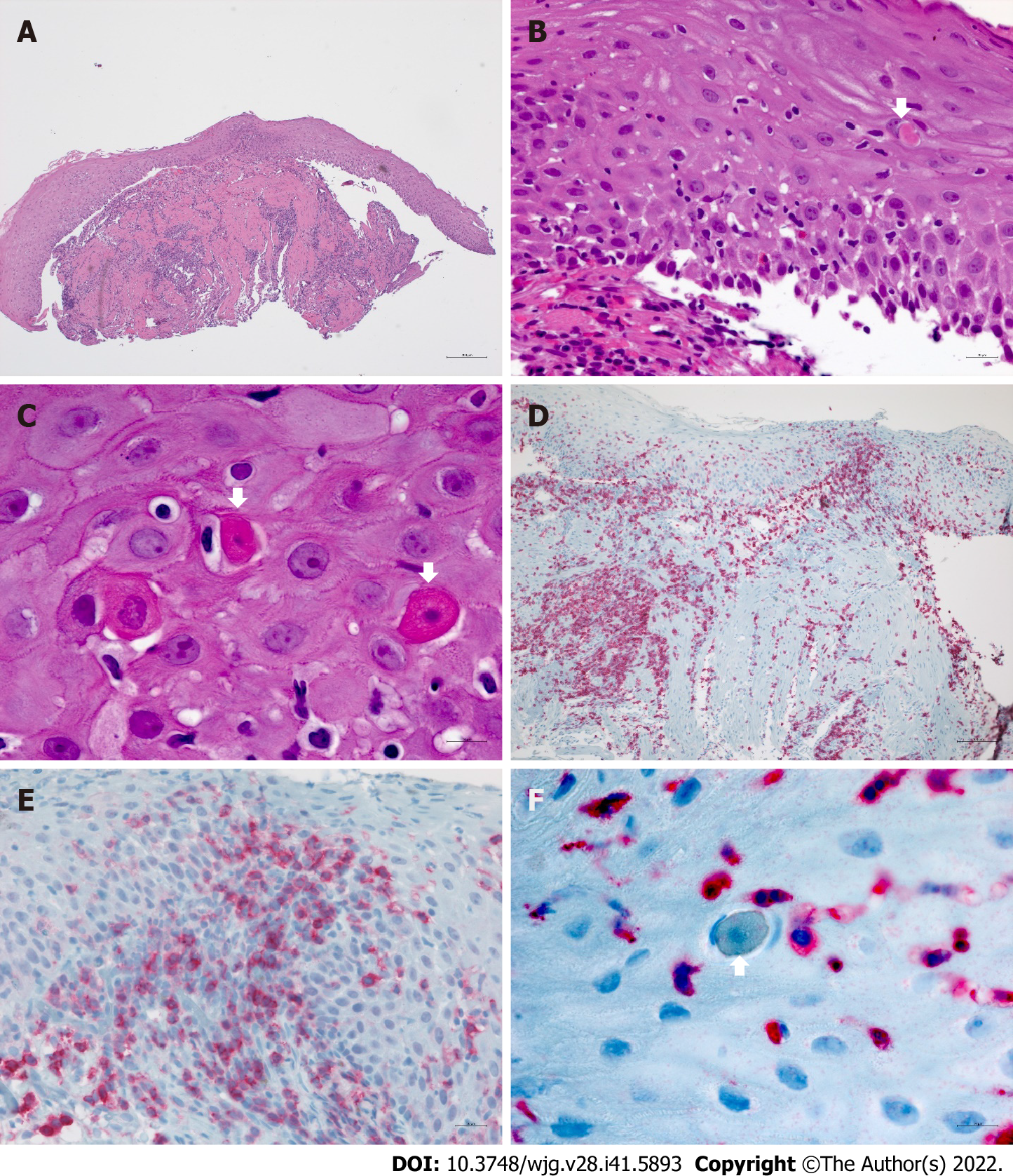
Figure 2 Histologic findings in esophageal lichen planus.
A and B: Lichenoid lymphocytic infiltrate of the lamina propria spilling over to the partially detached squamous epithelium; B and C: Intraepithelial lymphocytosis associated with apoptotic squamous cells (Civatte bodies, arrows); D: Dense CD3+ T-cell rich inflammation of the lamina propria involving 2/3 of surface epithelium and muscularis; E: Presence of a CD4+T-cell subset in the infiltrate; F: Civatte body rimmed by CD3+ T-cells.
- Citation: Decker A, Schauer F, Lazaro A, Monasterio C, Schmidt AR, Schmitt-Graeff A, Kreisel W. Esophageal lichen planus: Current knowledge, challenges and future perspectives. World J Gastroenterol 2022; 28(41): 5893-5909
- URL: https://www.wjgnet.com/1007-9327/full/v28/i41/5893.htm
- DOI: https://dx.doi.org/10.3748/wjg.v28.i41.5893


