Copyright
©The Author(s) 2022.
World J Gastroenterol. Nov 7, 2022; 28(41): 5893-5909
Published online Nov 7, 2022. doi: 10.3748/wjg.v28.i41.5893
Published online Nov 7, 2022. doi: 10.3748/wjg.v28.i41.5893
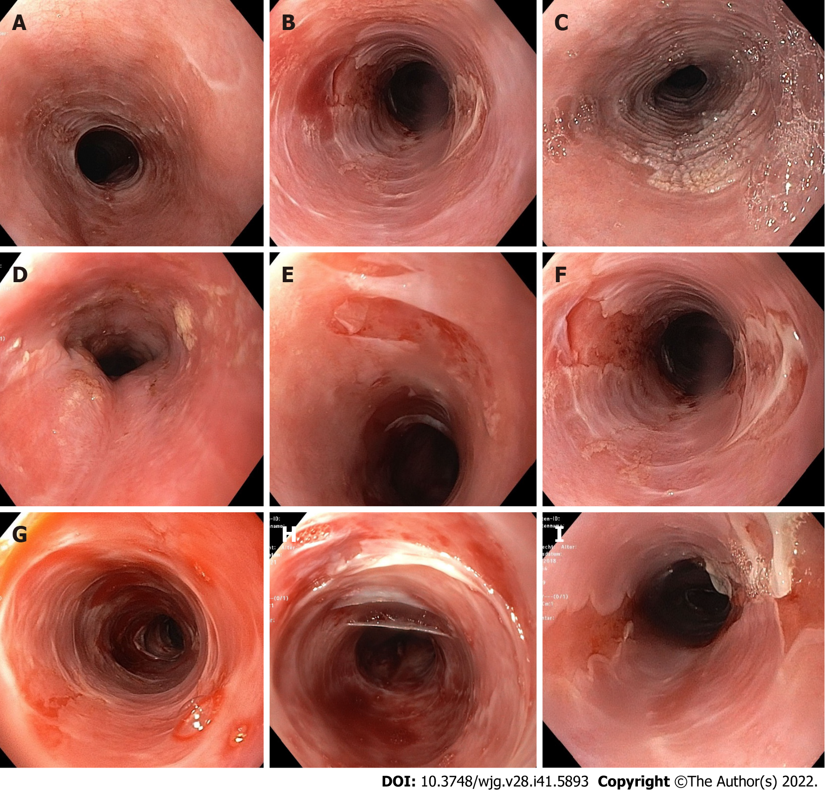
Figure 1 Endoscopic findings in esophageal lichen planus.
A: Trachealization; B: Trachealization and fragile mucosa; C: Hyperkeratosis; D: Hyperkeratosis and stenosis; E and F: Tearing and localized denudation of the mucosa; G-I: Tearing and spacious denudation of the mucosa. Endoscopic images were taken from our cohort of patients.
- Citation: Decker A, Schauer F, Lazaro A, Monasterio C, Schmidt AR, Schmitt-Graeff A, Kreisel W. Esophageal lichen planus: Current knowledge, challenges and future perspectives. World J Gastroenterol 2022; 28(41): 5893-5909
- URL: https://www.wjgnet.com/1007-9327/full/v28/i41/5893.htm
- DOI: https://dx.doi.org/10.3748/wjg.v28.i41.5893


