Copyright
©The Author(s) 2022.
World J Gastroenterol. Oct 28, 2022; 28(40): 5845-5864
Published online Oct 28, 2022. doi: 10.3748/wjg.v28.i40.5845
Published online Oct 28, 2022. doi: 10.3748/wjg.v28.i40.5845
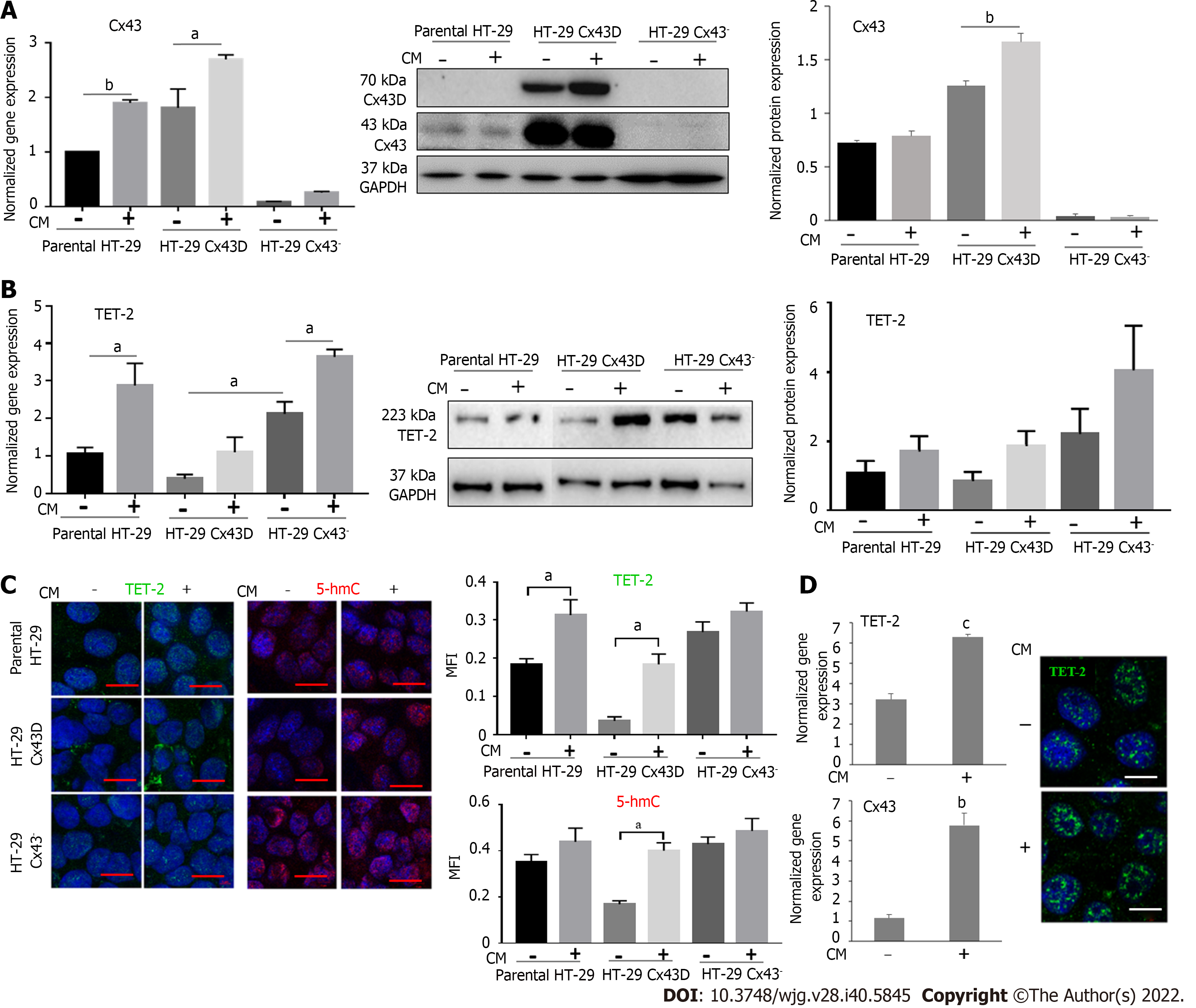
Figure 2 Connexin 43 and ten-eleven translocation-2 expression increases in cells exposed to inflammation.
Parental HT-29, HT-29 connexin 43-Dendra (Cx43D), and HT-29 Cx43- cells were exposed to inflammatory conditioned media (CM) obtained from activated THP-1 cells for 24 h. A: Histograms show the normalized gene expression of Cx43, as detected by quantitative polymerase chain reaction. Cx43 mRNA levels increase in parental HT-29 and HT-29 Cx43D, in the presence of inflammatory media. Western blot of endogenous Cx43 and exogenous Cx43D protein expression in parental HT-29, HT-29 Cx43D, and in HT-29 Cx43- cells. Densitometric analysis (ratio of protein-of-interest to loading control band intensity) shows a slight increase in Cx43 levels upon exposure to inflammation; B: Bar graphs show ten-eleven translocation-2 (TET-2) transcriptional levels increase in all three HT-29 cellular subsets when exposed to CM. Western blots of TET-2 and densitometric analysis shows increased protein levels of TET-2 in CM-treated cells; C: Immunofluorescence images showing TET-2 expression and 5-hmC marks in parental HT-29, HT-29 Cx43D, and HT-29 Cx43- cells. Bar graphs in the right panel reflect mean fluorescence intensity of at least five different fields acquired from three different experiments. Levels and activity of TET-2 increase in all CM-exposed cells. Scale bar 5 μm; D: Bar graphs display levels of Cx43 and TET-2 in Caco-2 cells exposed to CM. Fluorescent micrographs show increased levels of TET-2 in the nucleus upon exposure to CM. Scale bar 5 μm. Experiments were repeated at least three different times. One-way ANOVA, aP < 0.05; bP < 0.001; cP < 0.0005. TET-2: Translocation-2; Cx43: Connexin 43; GAPDH: Glyceraldehyde 3-phosphate dehydrogenase; MFI: Mean fluorescence intensity.
- Citation: El-Harakeh M, Saliba J, Sharaf Aldeen K, Haidar M, El Hajjar L, Awad MK, Hashash JG, Shirinian M, El-Sabban M. Expression of the methylcytosine dioxygenase ten-eleven translocation-2 and connexin 43 in inflammatory bowel disease and colorectal cancer. World J Gastroenterol 2022; 28(40): 5845-5864
- URL: https://www.wjgnet.com/1007-9327/full/v28/i40/5845.htm
- DOI: https://dx.doi.org/10.3748/wjg.v28.i40.5845


