Copyright
©The Author(s) 2022.
World J Gastroenterol. Oct 21, 2022; 28(39): 5707-5722
Published online Oct 21, 2022. doi: 10.3748/wjg.v28.i39.5707
Published online Oct 21, 2022. doi: 10.3748/wjg.v28.i39.5707
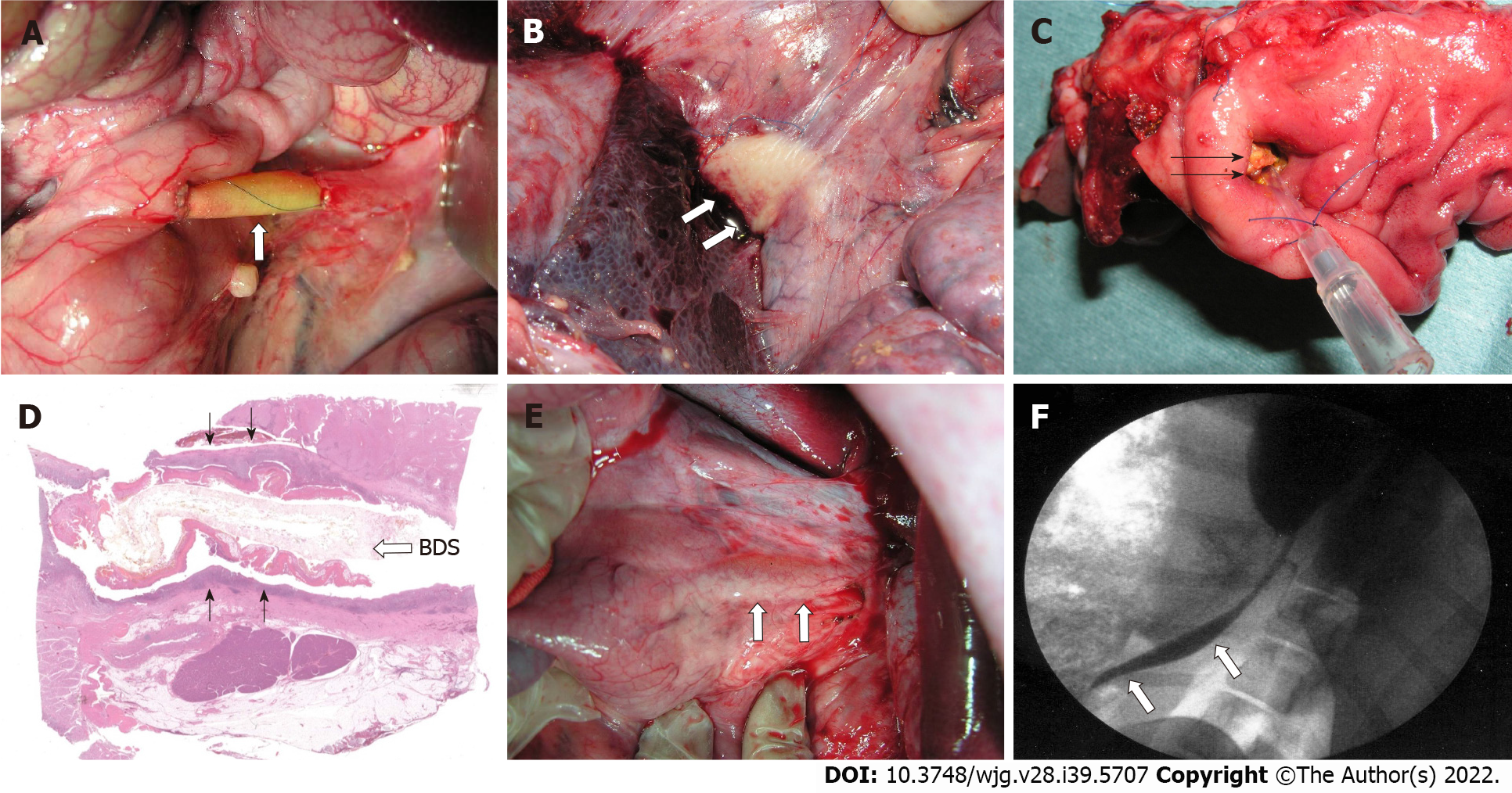
Figure 3 Bile duct substitutes using bioabsorbable material (synthetic polymer with short absorption period).
A: A bile duct substitute (BDS) using bioabsorbable material is implanted to bypass the extrahepatic bile duct (3 cm in size). Three weeks post-BDS implantation; B: White cell clusters are observed on the outside of the BDS; C: A vulnerable BDS is observed from the duodenal side; D: Dark purple connective tissue is noted on the outside of the BDS; E: At 6 mo after BDS implantation, the neo-bile duct is macroscopically similar to the natural common bile duct have been regenerated (arrow); F: Cholangiography (6 mo after BDS implantation). The BDS implant and anastomotic site become unknown, and the contrast medium flows smoothly into the duodenum (white arrows).
- Citation: Miyazawa M, Aikawa M, Takashima J, Kobayashi H, Ohnishi S, Ikada Y. Pitfalls and promises of bile duct alternatives: A narrative review. World J Gastroenterol 2022; 28(39): 5707-5722
- URL: https://www.wjgnet.com/1007-9327/full/v28/i39/5707.htm
- DOI: https://dx.doi.org/10.3748/wjg.v28.i39.5707


