Copyright
©The Author(s) 2022.
World J Gastroenterol. Sep 21, 2022; 28(35): 5237-5239
Published online Sep 21, 2022. doi: 10.3748/wjg.v28.i35.5237
Published online Sep 21, 2022. doi: 10.3748/wjg.v28.i35.5237
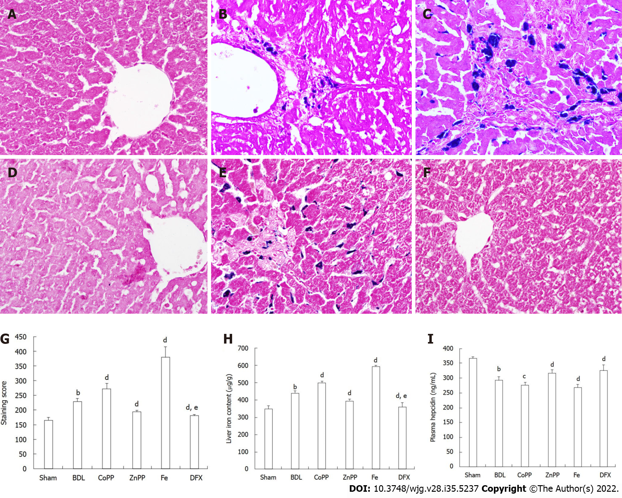
Figure 1 erl’s Prussian blue staining, levels of hepcidin, serum and liver iron.
A: No iron accumulated in the Sham group; B: A small amount of iron mainly accumulated on Kupffer cells in the bile duct ligation (BDL) group; C: Much more iron accumulation was found in interlobular and macrophagocytes in the cobalt protoporphyrin (CoPP) group; D and F: Almost no iron accumulation was detected in the zinc protoporphyrin (ZnPP) group and deferoxamine (DFX) group; E: Massive iron accumulation was observed in the Fe group; G and H: There were no differences in the hepatic and serum iron content of these six groups; I: Plasma hepcidin also was measured by enzyme-linked immuno sorbent assay (magnification × 400). Values are expressed as mean ± SE (n = 6). bP < 0.01 vs Sham group; cP < 0.05, dP < 0.01 vs BDL group; eP < 0.05 vs ZnPP group.
- Citation: Wang QM, Du JL, Duan ZJ, Guo SB, Sun XY, Liu Z. Correction to “Inhibiting heme oxygenase-1 attenuates rat liver fibrosis by removing iron accumulation”. World J Gastroenterol 2022; 28(35): 5237-5239
- URL: https://www.wjgnet.com/1007-9327/full/v28/i35/5237.htm
- DOI: https://dx.doi.org/10.3748/wjg.v28.i35.5237


