Copyright
©The Author(s) 2022.
World J Gastroenterol. Sep 14, 2022; 28(34): 5047-5057
Published online Sep 14, 2022. doi: 10.3748/wjg.v28.i34.5047
Published online Sep 14, 2022. doi: 10.3748/wjg.v28.i34.5047
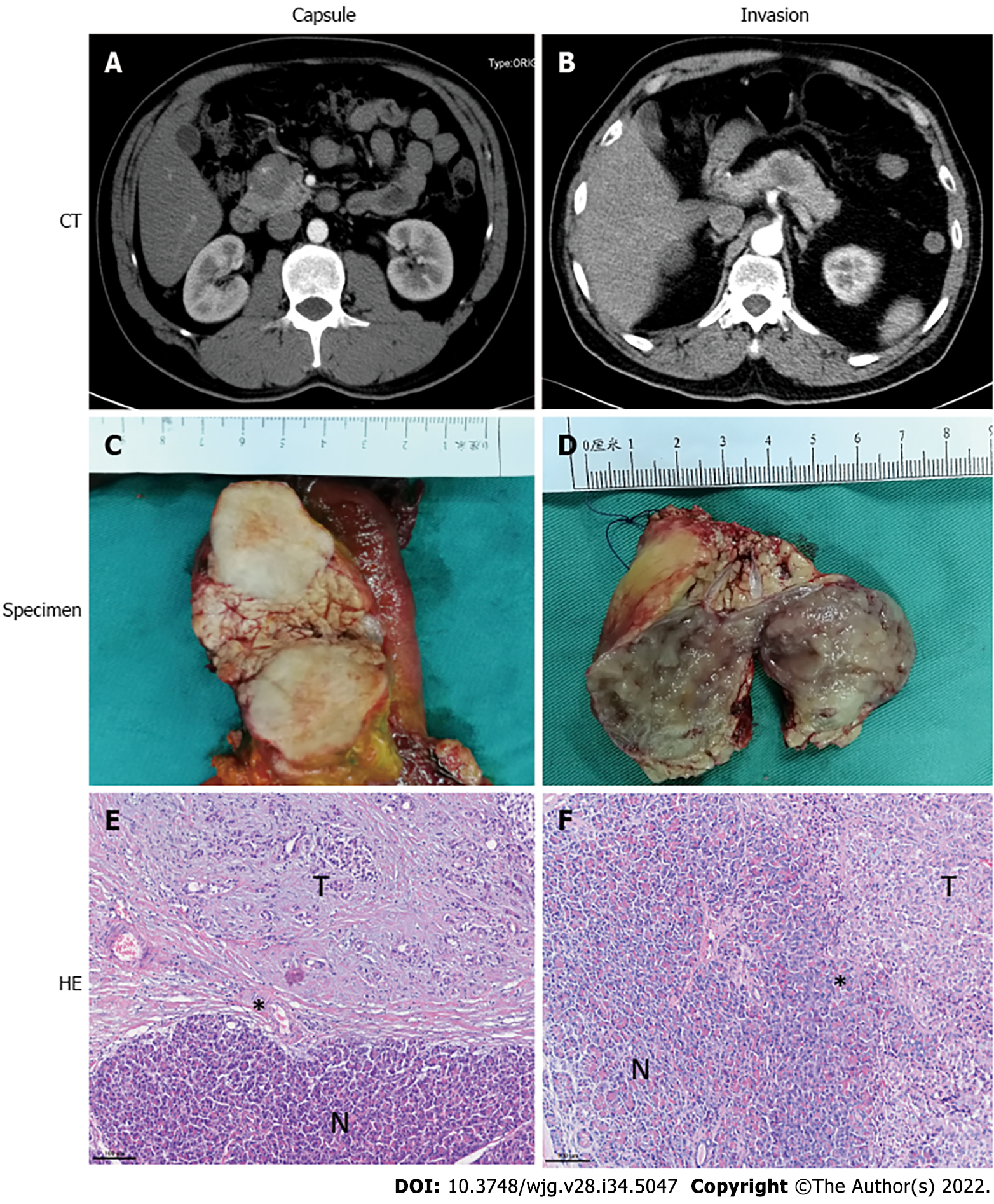
Figure 1 Computed tomography imaging, specimen and histopathologic features of capsulized and invasive solid pseudopapillary tumors.
A, C and E: For the capsulized solid pseudopapillary tumors (SPTs), computed tomography (CT) imaging (A) and specimen (C) showed a clear distinction from surrounding pancreatic tissue, and an intact fibrous envelope on the tumor margin on hematoxylin and eosin (HE)-stained section (E); B, D and F: Invasive SPT was indistinguishable from surrounding normal pancreatic tissue on CT imaging (B) and in the specimen (D), and tumor cells showed infiltrative growth into normal pancreatic tissue on HE-stained section (F). N: Normal pancreas tissue; T: Tumor. *Tumor margin.
- Citation: Yang J, Tan CL, Long D, Liang Y, Zhou L, Liu XB, Chen YH. Analysis of invasiveness and tumor-associated macrophages infiltration in solid pseudopapillary tumors of pancreas. World J Gastroenterol 2022; 28(34): 5047-5057
- URL: https://www.wjgnet.com/1007-9327/full/v28/i34/5047.htm
- DOI: https://dx.doi.org/10.3748/wjg.v28.i34.5047


