Copyright
©The Author(s) 2022.
World J Gastroenterol. Sep 7, 2022; 28(33): 4920-4925
Published online Sep 7, 2022. doi: 10.3748/wjg.v28.i33.4920
Published online Sep 7, 2022. doi: 10.3748/wjg.v28.i33.4920
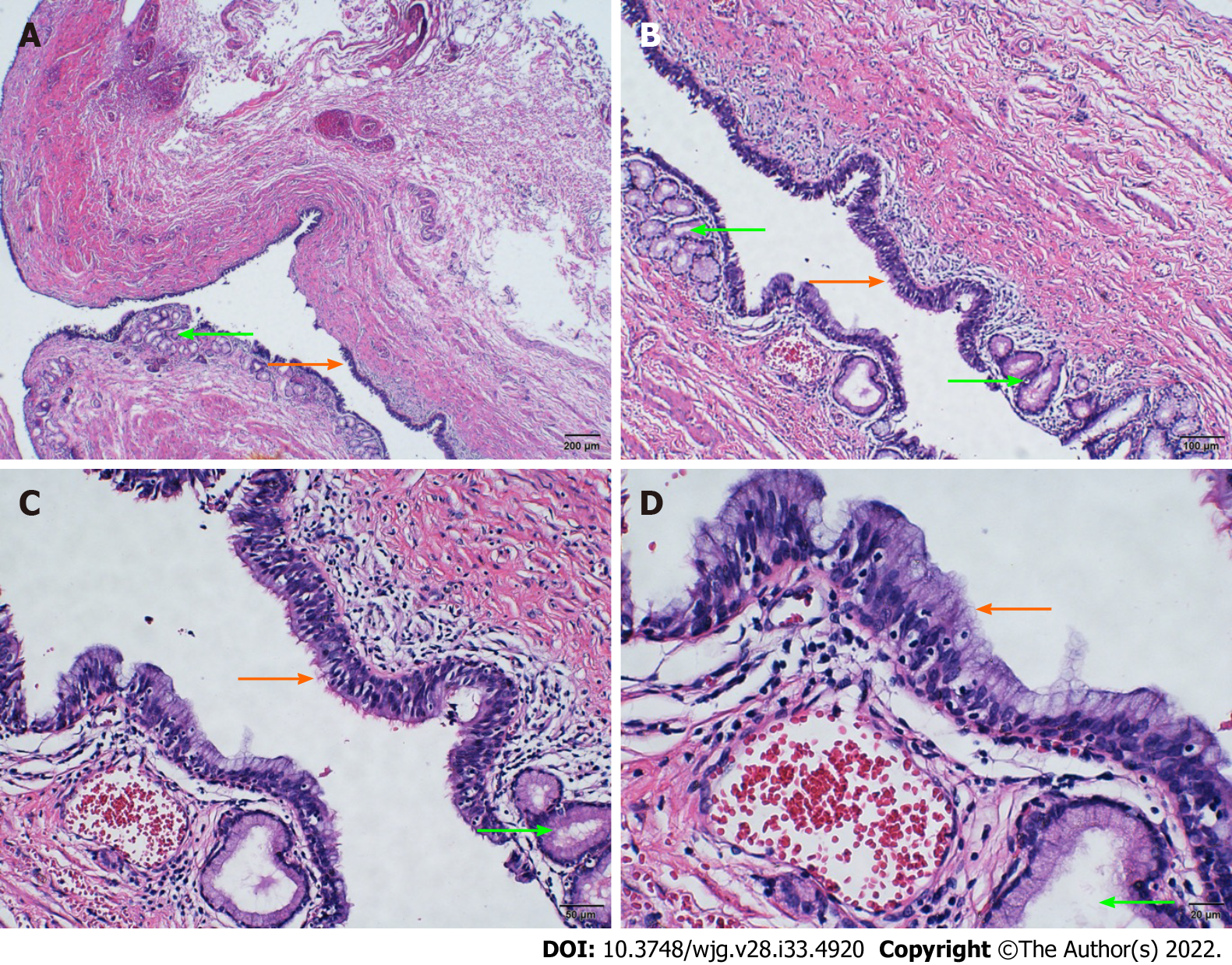
Figure 3 Histopathologic examination by hematoxylin and eosin staining.
The cyst wall was fibrous and muscle tissue was visible. The mucosal epithelium of the inner surface of the cyst wall was partially detached, and the remaining inner surface was partly lined with pseudostratified ciliated columnar epithelium (orange arrow) and mucous epithelium. Serous and mucous glands were observed in the lamina propria (green arrow). A: 4 ×; B: 10 ×; C: 20 ×; D: 40 ×.
- Citation: Dong CJ, Yang RM, Wang QL, Wu QY, Yang DJ, Kong DC, Zhang P. Ectopic bronchogenic cyst of liver misdiagnosed as gallbladder diverticulum: A case report. World J Gastroenterol 2022; 28(33): 4920-4925
- URL: https://www.wjgnet.com/1007-9327/full/v28/i33/4920.htm
- DOI: https://dx.doi.org/10.3748/wjg.v28.i33.4920


