Copyright
©The Author(s) 2022.
World J Gastroenterol. Sep 7, 2022; 28(33): 4920-4925
Published online Sep 7, 2022. doi: 10.3748/wjg.v28.i33.4920
Published online Sep 7, 2022. doi: 10.3748/wjg.v28.i33.4920
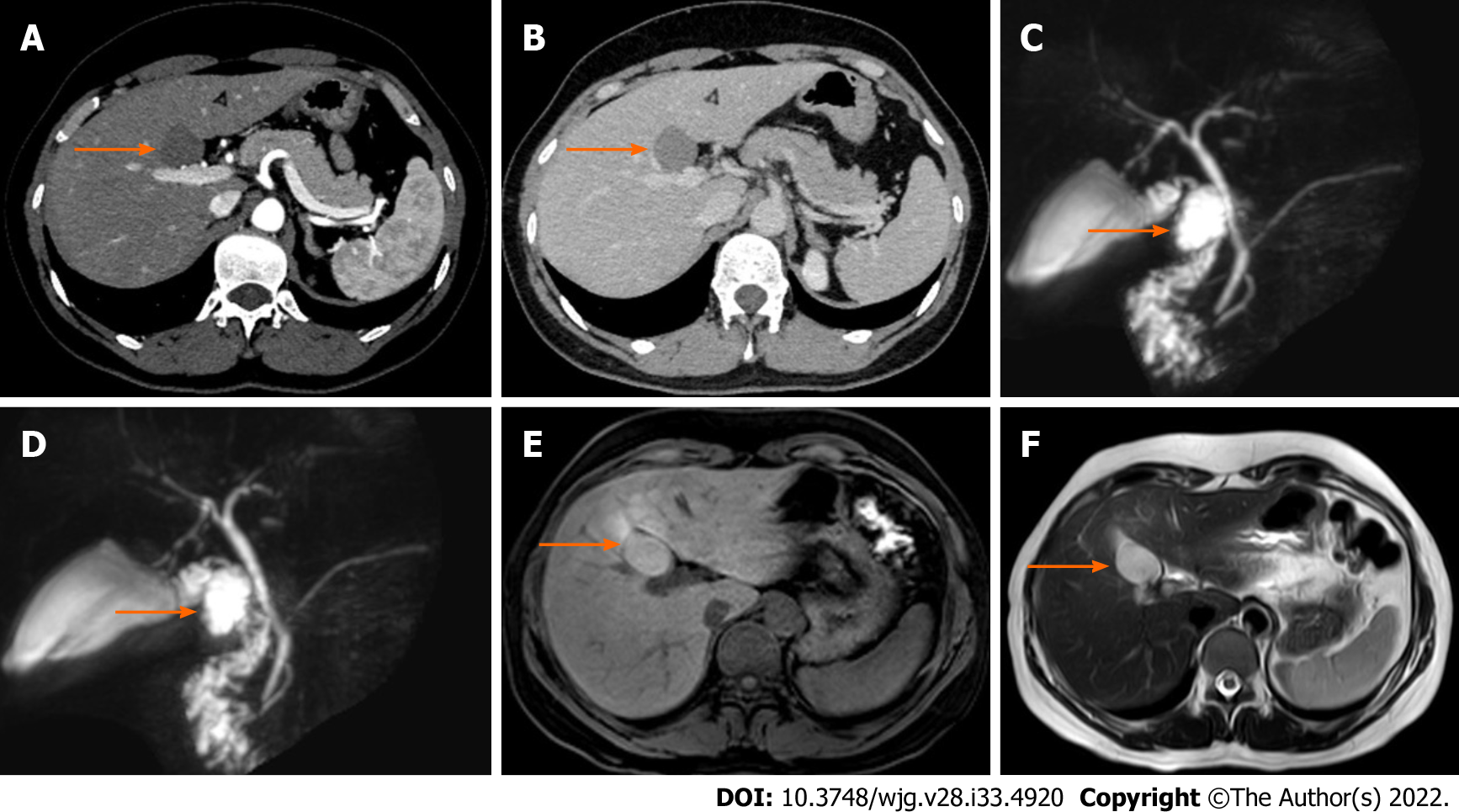
Figure 1 Ectopic bronchogenic cyst in a 40-year-old woman.
The location of the cyst is indicated by an orange arrow. Magnetic resonance cholangiopancreatography (MRCP) showed that the cyst was locally connected to the cystic duct. A: Hepatobiliary and pancreatic enhanced computed tomography (CT) arterial phase; B: Hepatobiliary and pancreatic enhanced CT venous phase; C and D: MRCP; E: T1-weighted imaging showed a slightly high signal shadow; F: T2-weighted imaging showed a high signal shadow.
- Citation: Dong CJ, Yang RM, Wang QL, Wu QY, Yang DJ, Kong DC, Zhang P. Ectopic bronchogenic cyst of liver misdiagnosed as gallbladder diverticulum: A case report. World J Gastroenterol 2022; 28(33): 4920-4925
- URL: https://www.wjgnet.com/1007-9327/full/v28/i33/4920.htm
- DOI: https://dx.doi.org/10.3748/wjg.v28.i33.4920


