Copyright
©The Author(s) 2022.
World J Gastroenterol. Aug 28, 2022; 28(32): 4540-4556
Published online Aug 28, 2022. doi: 10.3748/wjg.v28.i32.4540
Published online Aug 28, 2022. doi: 10.3748/wjg.v28.i32.4540
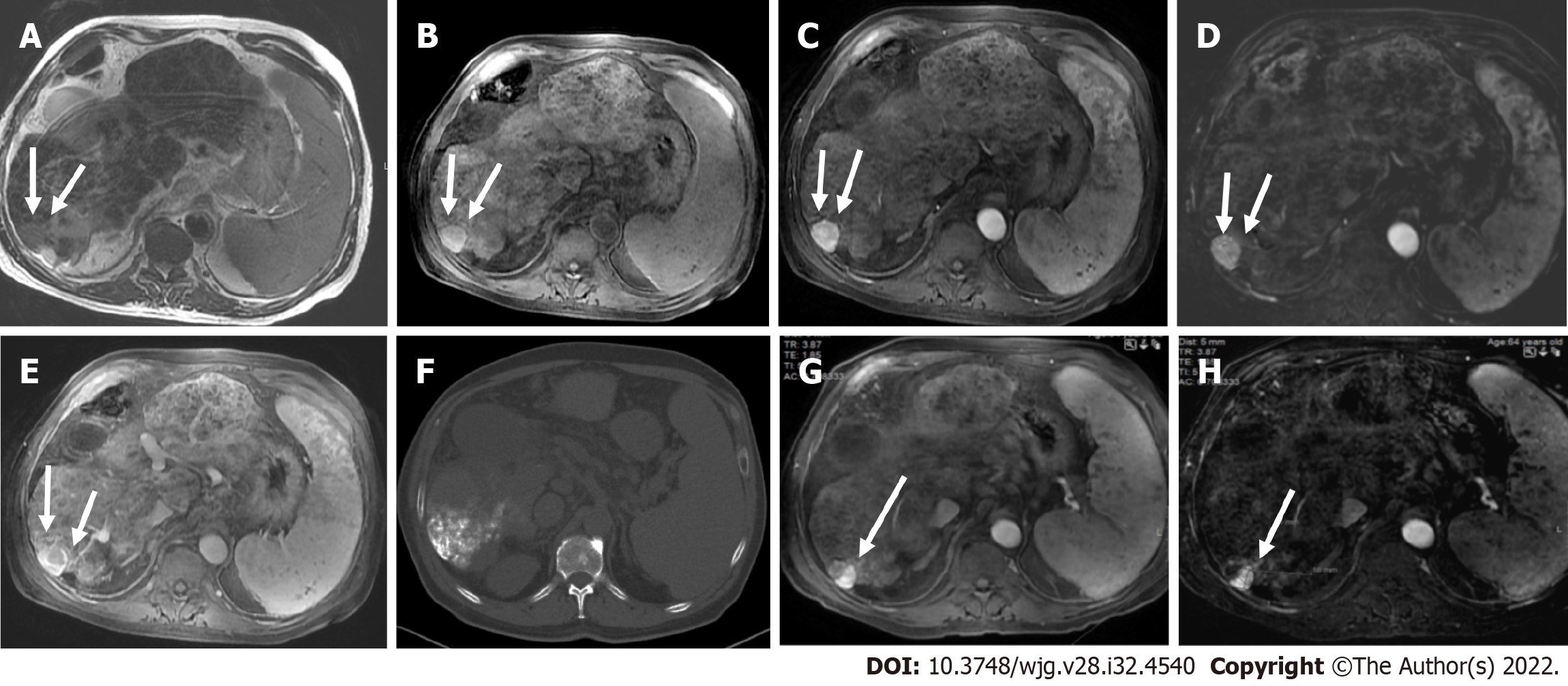
Figure 18 LR-5 observation.
A and B: Axial T2 weighted (A) and volumetric interpolated breath-hold examination (B) show a segment VI observation with high signal intensity (arrows); C and D: On arterial phase (C) strong enhancement is seen, better depicted on subtracted image (D); E: On portal phase washout is seen categorized as LR-5; F: Unenhanced computed tomography scan after TACE shows accumulation of iodized oil (arrow) within the tumor; G and H: However on post-contrast follow up magnetic resonance imaging on arterial phase (G) there is residual nodular enhancement within a lesion clearly depicted on subtracted image (H) categorized as LR-treatment response viable.
- Citation: Liava C, Sinakos E, Papadopoulou E, Giannakopoulou L, Potsi S, Moumtzouoglou A, Chatziioannou A, Stergioulas L, Kalogeropoulou L, Dedes I, Akriviadis E, Chourmouzi D. Liver Imaging Reporting and Data System criteria for the diagnosis of hepatocellular carcinoma in clinical practice: A pictorial minireview. World J Gastroenterol 2022; 28(32): 4540-4556
- URL: https://www.wjgnet.com/1007-9327/full/v28/i32/4540.htm
- DOI: https://dx.doi.org/10.3748/wjg.v28.i32.4540


