Copyright
©The Author(s) 2022.
World J Gastroenterol. Aug 28, 2022; 28(32): 4540-4556
Published online Aug 28, 2022. doi: 10.3748/wjg.v28.i32.4540
Published online Aug 28, 2022. doi: 10.3748/wjg.v28.i32.4540
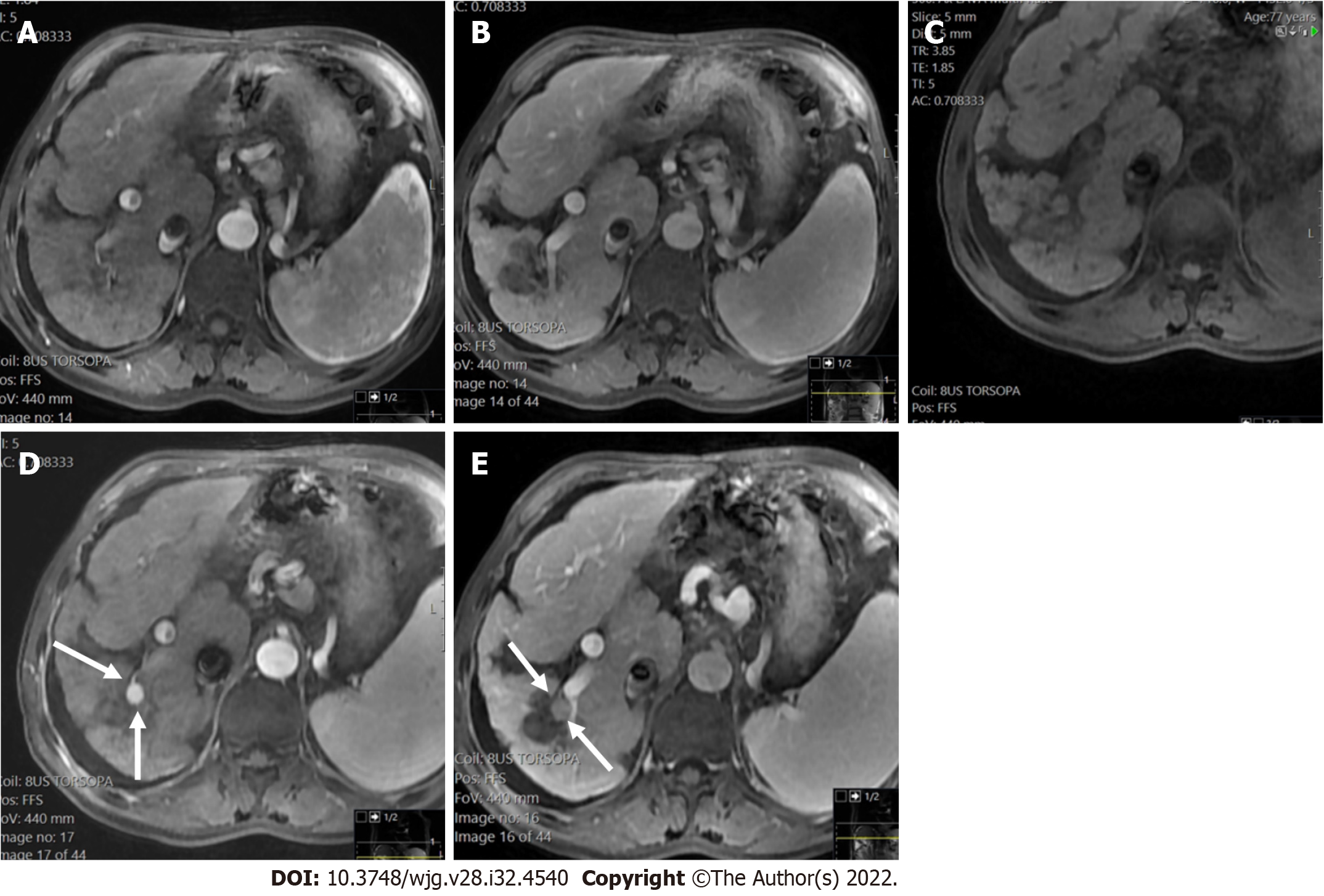
Figure 17 LR-5 observation.
A and B: Axial post-contrast images arterial (A) and portal phase (B) showed an ablation defect in segment VI without residual areas of arterial enhancement or washout. This was classified as LR-treatment response non-viable; C-E: Later pre-contrast (C) arterial (D) and portal (E) phase showed a new nodule (arrows) measuring 12 mm with strong arterial enhancement and washout categorized as LR-5. If a new tumor arises within or adjacent to a treated lesion, this observation should be assigned a new non-treated LI-RADS category.
- Citation: Liava C, Sinakos E, Papadopoulou E, Giannakopoulou L, Potsi S, Moumtzouoglou A, Chatziioannou A, Stergioulas L, Kalogeropoulou L, Dedes I, Akriviadis E, Chourmouzi D. Liver Imaging Reporting and Data System criteria for the diagnosis of hepatocellular carcinoma in clinical practice: A pictorial minireview. World J Gastroenterol 2022; 28(32): 4540-4556
- URL: https://www.wjgnet.com/1007-9327/full/v28/i32/4540.htm
- DOI: https://dx.doi.org/10.3748/wjg.v28.i32.4540


