Copyright
©The Author(s) 2022.
World J Gastroenterol. Aug 28, 2022; 28(32): 4540-4556
Published online Aug 28, 2022. doi: 10.3748/wjg.v28.i32.4540
Published online Aug 28, 2022. doi: 10.3748/wjg.v28.i32.4540
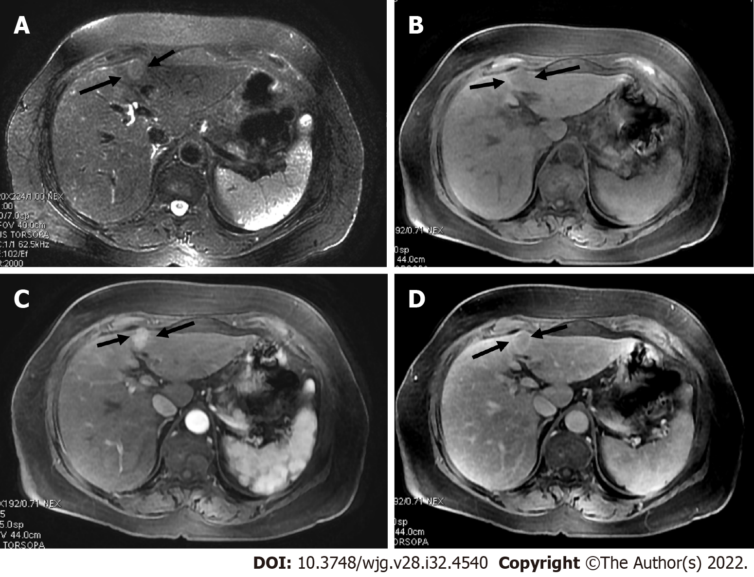
Figure 14 A 62-year-old female with chronic hepatitis C.
A: On axial short tau inversion recovery image, a 21 mm IVb segment observation with relative high signal intensity is seen (arrows); B: On axial volumetric interpolated breath-hold examination image the observation shows low signal intensity (arrows); C and D: On post-contrast images, major features of hepatocellular carcinoma (HCC), arterial phase hyperenhancement (arrows) on arterial phase (C), washout and enhancing capsule (arrows) on portal (D) were seen, indicating LR-5 (definitely HCC).
- Citation: Liava C, Sinakos E, Papadopoulou E, Giannakopoulou L, Potsi S, Moumtzouoglou A, Chatziioannou A, Stergioulas L, Kalogeropoulou L, Dedes I, Akriviadis E, Chourmouzi D. Liver Imaging Reporting and Data System criteria for the diagnosis of hepatocellular carcinoma in clinical practice: A pictorial minireview. World J Gastroenterol 2022; 28(32): 4540-4556
- URL: https://www.wjgnet.com/1007-9327/full/v28/i32/4540.htm
- DOI: https://dx.doi.org/10.3748/wjg.v28.i32.4540


