Copyright
©The Author(s) 2022.
World J Gastroenterol. Aug 28, 2022; 28(32): 4540-4556
Published online Aug 28, 2022. doi: 10.3748/wjg.v28.i32.4540
Published online Aug 28, 2022. doi: 10.3748/wjg.v28.i32.4540
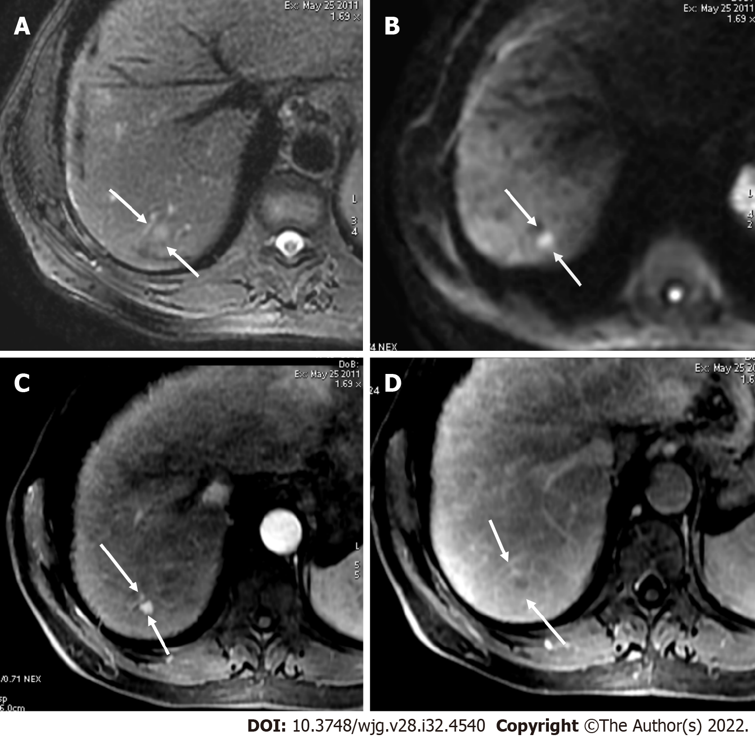
Figure 13 Axial short tau inversion recovery and diffusion-weighted imaging.
A and B: Axial short tau inversion recovery (A) and diffusion-weighted imaging (DWI) (B) images showed a < 10 mm segment VII observation with high signal intensity; C and D: In the arterial phase (C), arterial phase hyperenhancement was seen with washout in the portal phase (D) categorized as LR-4. Restriction on DWI does not allow to upgrade from LR-4 to LR-5, because ancillary features lack sufficient specificity for hepatocellular carcinoma to allow for an LR-5 upgrade.
- Citation: Liava C, Sinakos E, Papadopoulou E, Giannakopoulou L, Potsi S, Moumtzouoglou A, Chatziioannou A, Stergioulas L, Kalogeropoulou L, Dedes I, Akriviadis E, Chourmouzi D. Liver Imaging Reporting and Data System criteria for the diagnosis of hepatocellular carcinoma in clinical practice: A pictorial minireview. World J Gastroenterol 2022; 28(32): 4540-4556
- URL: https://www.wjgnet.com/1007-9327/full/v28/i32/4540.htm
- DOI: https://dx.doi.org/10.3748/wjg.v28.i32.4540


