Copyright
©The Author(s) 2022.
World J Gastroenterol. Aug 28, 2022; 28(32): 4540-4556
Published online Aug 28, 2022. doi: 10.3748/wjg.v28.i32.4540
Published online Aug 28, 2022. doi: 10.3748/wjg.v28.i32.4540
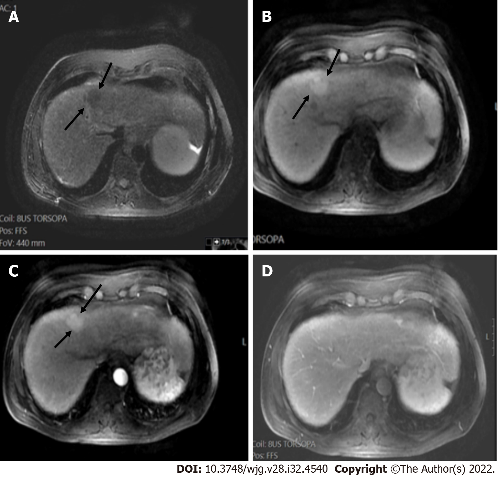
Figure 12 Axial short tau inversion recovery image.
A and B: A IVa segment observation with low signal intensity (arrows) measuring > 20 mm (A) and high signal intensity on pre-contrast image (arrows on B); C and D: On arterial (C) and portal (D) phase there was no arterial phase hyperenhancement or washout and the lesion was categorized as LR-3.
- Citation: Liava C, Sinakos E, Papadopoulou E, Giannakopoulou L, Potsi S, Moumtzouoglou A, Chatziioannou A, Stergioulas L, Kalogeropoulou L, Dedes I, Akriviadis E, Chourmouzi D. Liver Imaging Reporting and Data System criteria for the diagnosis of hepatocellular carcinoma in clinical practice: A pictorial minireview. World J Gastroenterol 2022; 28(32): 4540-4556
- URL: https://www.wjgnet.com/1007-9327/full/v28/i32/4540.htm
- DOI: https://dx.doi.org/10.3748/wjg.v28.i32.4540


