Copyright
©The Author(s) 2022.
World J Gastroenterol. Aug 28, 2022; 28(32): 4540-4556
Published online Aug 28, 2022. doi: 10.3748/wjg.v28.i32.4540
Published online Aug 28, 2022. doi: 10.3748/wjg.v28.i32.4540
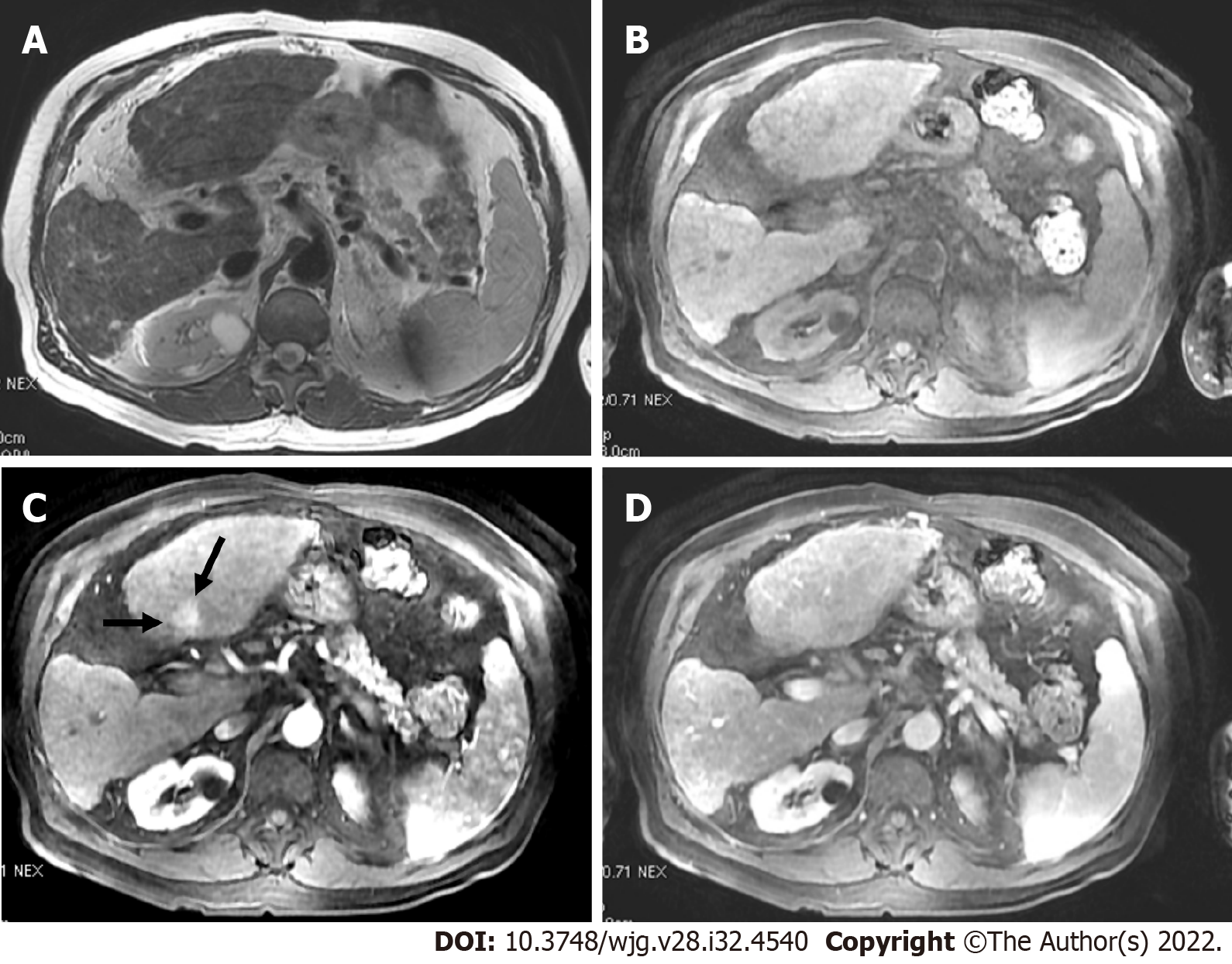
Figure 11 T2 weighted image and volumetric interpolated breath-hold examination.
A and B: Axial T2 weighted image (A) and volumetric interpolated breath-hold examination (VIBE) (B) showed a shrunken and nodular liver that was consistent with cirrhosis; C and D: On post-contrast VIBE arterial-phase image (C), a 1.6 cm lesion in segment IVb demonstrated hyperenhancement (arrows) without washout on portal venous phase image (D). According to Liver Imaging Reporting and Data System criteria, the lesion was categorized as LR-3.
- Citation: Liava C, Sinakos E, Papadopoulou E, Giannakopoulou L, Potsi S, Moumtzouoglou A, Chatziioannou A, Stergioulas L, Kalogeropoulou L, Dedes I, Akriviadis E, Chourmouzi D. Liver Imaging Reporting and Data System criteria for the diagnosis of hepatocellular carcinoma in clinical practice: A pictorial minireview. World J Gastroenterol 2022; 28(32): 4540-4556
- URL: https://www.wjgnet.com/1007-9327/full/v28/i32/4540.htm
- DOI: https://dx.doi.org/10.3748/wjg.v28.i32.4540


