Copyright
©The Author(s) 2022.
World J Gastroenterol. Aug 28, 2022; 28(32): 4540-4556
Published online Aug 28, 2022. doi: 10.3748/wjg.v28.i32.4540
Published online Aug 28, 2022. doi: 10.3748/wjg.v28.i32.4540
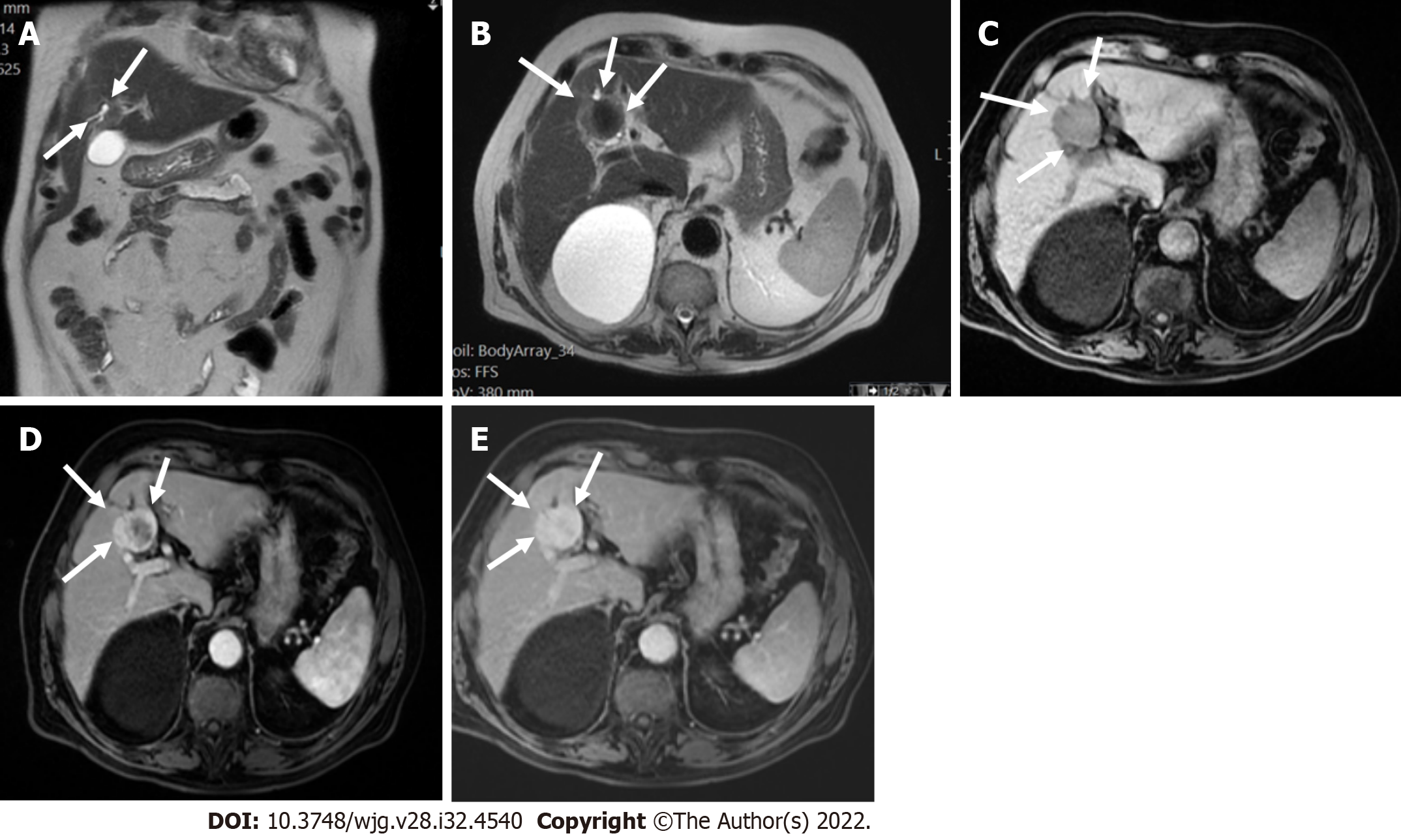
Figure 10 T1 weighted image and T2 weighted image.
A and B: Coronal (A) and axial (B) T2 weighted images showed a low signal segment V mass in a patient with chronic hepatitis B (arrows); C: The mass showed low signal intensity (arrows) on T1 weighted image; D and E: In the arterial phase (D), there was peripheral enhancement (arrows) and progressive filling (arrows) in the portal phase (E) categorized as LR-M. Histologic examination confirmed the diagnosis of cholangiocarcinoma and not hepatocellular carcinoma.
- Citation: Liava C, Sinakos E, Papadopoulou E, Giannakopoulou L, Potsi S, Moumtzouoglou A, Chatziioannou A, Stergioulas L, Kalogeropoulou L, Dedes I, Akriviadis E, Chourmouzi D. Liver Imaging Reporting and Data System criteria for the diagnosis of hepatocellular carcinoma in clinical practice: A pictorial minireview. World J Gastroenterol 2022; 28(32): 4540-4556
- URL: https://www.wjgnet.com/1007-9327/full/v28/i32/4540.htm
- DOI: https://dx.doi.org/10.3748/wjg.v28.i32.4540


