Copyright
©The Author(s) 2022.
World J Gastroenterol. Aug 28, 2022; 28(32): 4540-4556
Published online Aug 28, 2022. doi: 10.3748/wjg.v28.i32.4540
Published online Aug 28, 2022. doi: 10.3748/wjg.v28.i32.4540
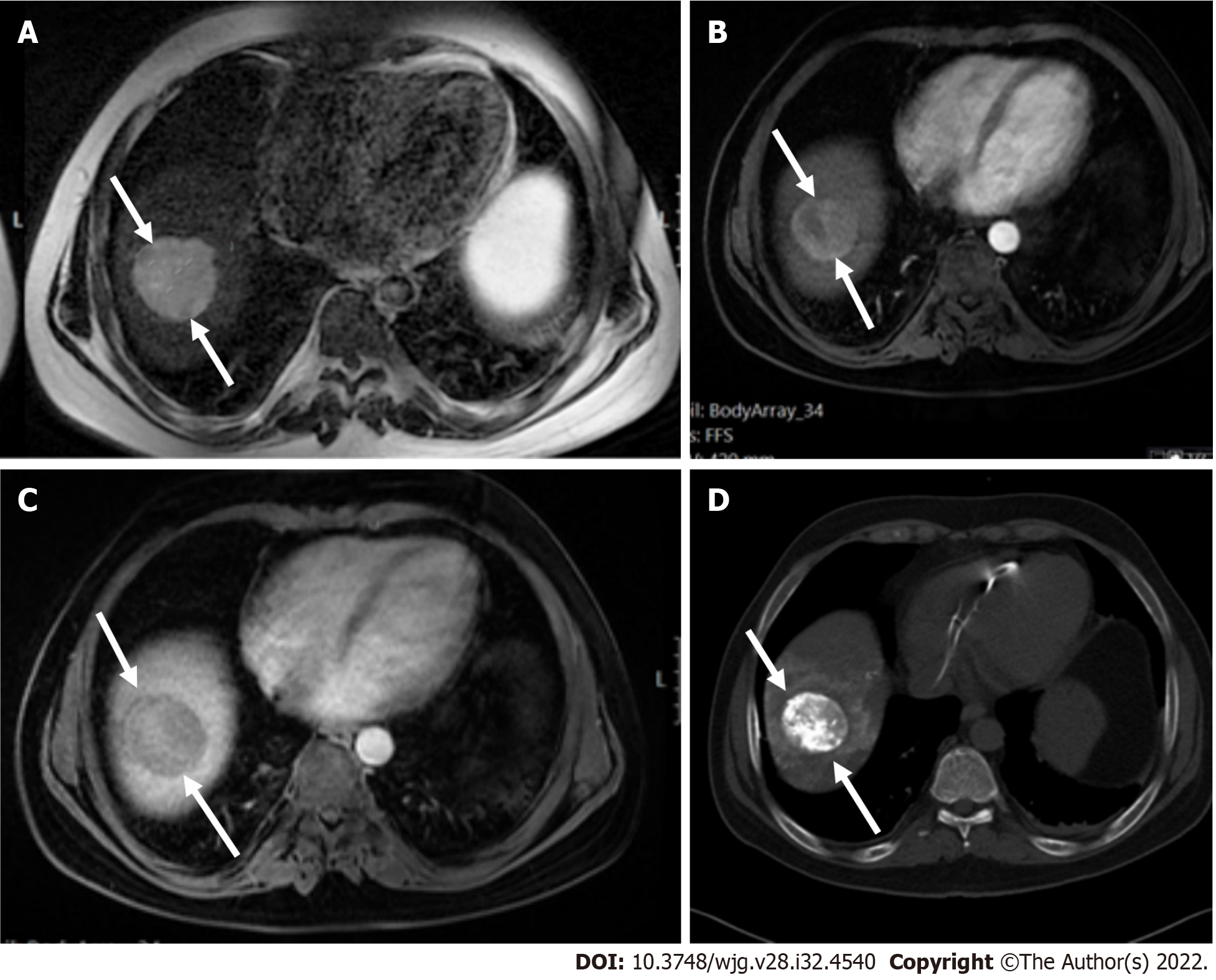
Figure 3 A 47 mm liver mass was incidentally found, in a 40-year-old man with beta thalassemia, during cardiac magnetic resonance investigation for myocardial iron overload.
A-C: On abdominal magnetic resonance imaging, the lesion demonstrated high signal intensity on T2 weighted image (A), arterial phase hyperenhancement on post-contrast arterial phase (B), and washout on portal phase (C). Although there are no current evidence-based guidelines for patients with beta thalassemia without cirrhosis, the hepatologist confirmed the patient’s risk status and proposed that Liver Imaging Reporting and Data System (LI-RADS) should be applied. According to the LI-RADS criteria, the lesion was categorized as LR-5 (definite hepatocellular carcinoma); D: Unenhanced computed tomography scan after transarterial chemoembolization showed accumulation of iodized oil (arrow) within the tumor.
- Citation: Liava C, Sinakos E, Papadopoulou E, Giannakopoulou L, Potsi S, Moumtzouoglou A, Chatziioannou A, Stergioulas L, Kalogeropoulou L, Dedes I, Akriviadis E, Chourmouzi D. Liver Imaging Reporting and Data System criteria for the diagnosis of hepatocellular carcinoma in clinical practice: A pictorial minireview. World J Gastroenterol 2022; 28(32): 4540-4556
- URL: https://www.wjgnet.com/1007-9327/full/v28/i32/4540.htm
- DOI: https://dx.doi.org/10.3748/wjg.v28.i32.4540


