Copyright
©The Author(s) 2022.
World J Gastroenterol. Aug 28, 2022; 28(32): 4540-4556
Published online Aug 28, 2022. doi: 10.3748/wjg.v28.i32.4540
Published online Aug 28, 2022. doi: 10.3748/wjg.v28.i32.4540
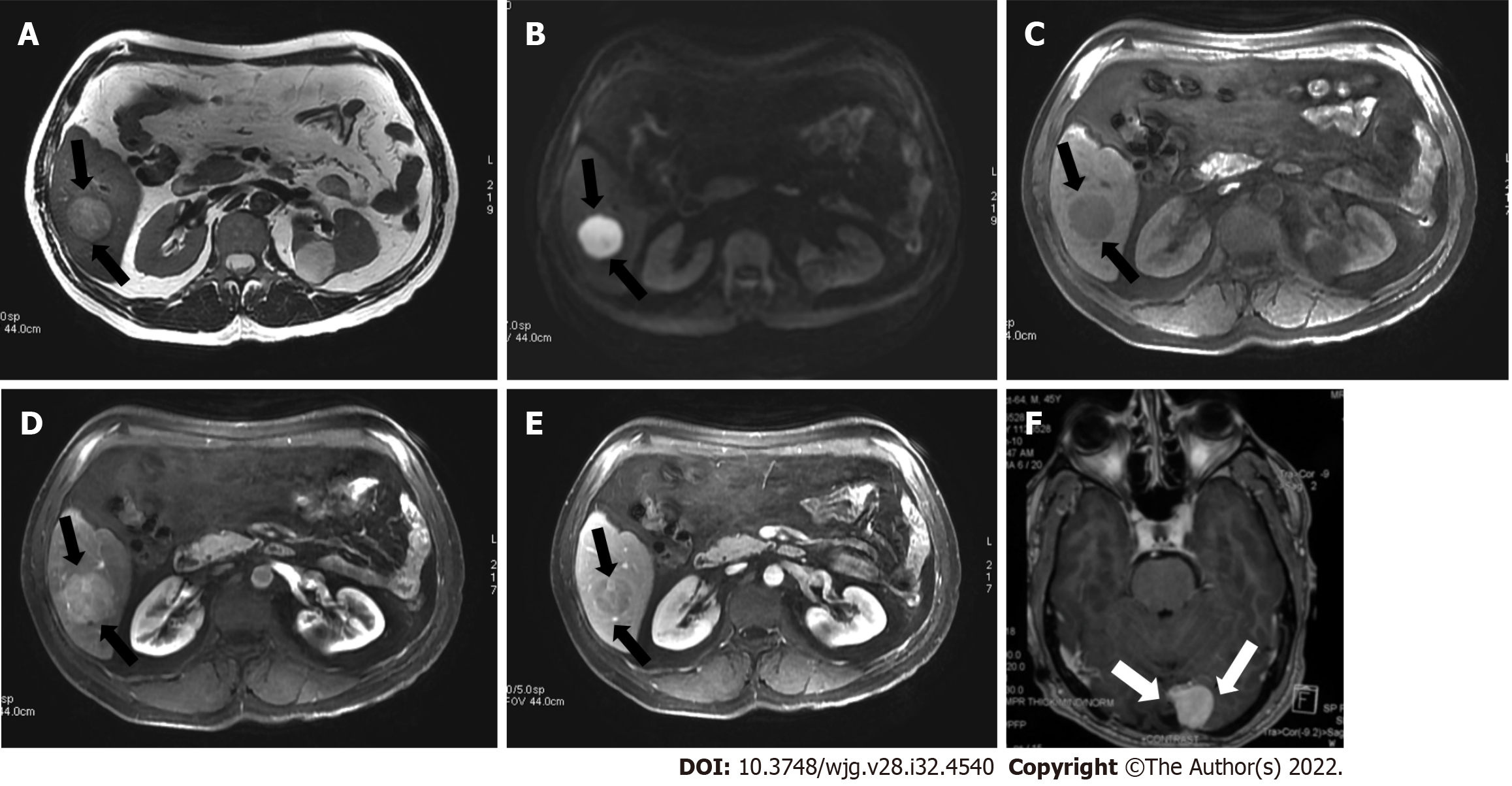
Figure 2 A 45-year-old man with a segment VI lesion.
A: The lesion demonstrated a high signal on T2 weighted image; B: High on diffusion-weighted imaging; C: Low on volumetric interpolated breath-hold examination; D: Non-rim arterial-phase hyper-enhancement on arterial-phase image; E: Washout on portal phase. If we applied Liver Imaging Reporting and Data System (LI-RADS) criteria, the erroneous diagnosis of hepatocellular carcinoma could be made. However, the patient did not meet the at-risk criteria to apply LI-RADS. Histologic examination of the lesion demonstrated metastatic hemangiopericytoma; F: Magnetic resonance imaging of the brain, axial post-contrast T1-weighted image showed an extra-axial lobulated, occipital enhancing mass corresponding to recurrence of hemangiopericytoma 12 mo after total surgical excision.
- Citation: Liava C, Sinakos E, Papadopoulou E, Giannakopoulou L, Potsi S, Moumtzouoglou A, Chatziioannou A, Stergioulas L, Kalogeropoulou L, Dedes I, Akriviadis E, Chourmouzi D. Liver Imaging Reporting and Data System criteria for the diagnosis of hepatocellular carcinoma in clinical practice: A pictorial minireview. World J Gastroenterol 2022; 28(32): 4540-4556
- URL: https://www.wjgnet.com/1007-9327/full/v28/i32/4540.htm
- DOI: https://dx.doi.org/10.3748/wjg.v28.i32.4540


