Copyright
©The Author(s) 2022.
World J Gastroenterol. Aug 21, 2022; 28(31): 4363-4375
Published online Aug 21, 2022. doi: 10.3748/wjg.v28.i31.4363
Published online Aug 21, 2022. doi: 10.3748/wjg.v28.i31.4363
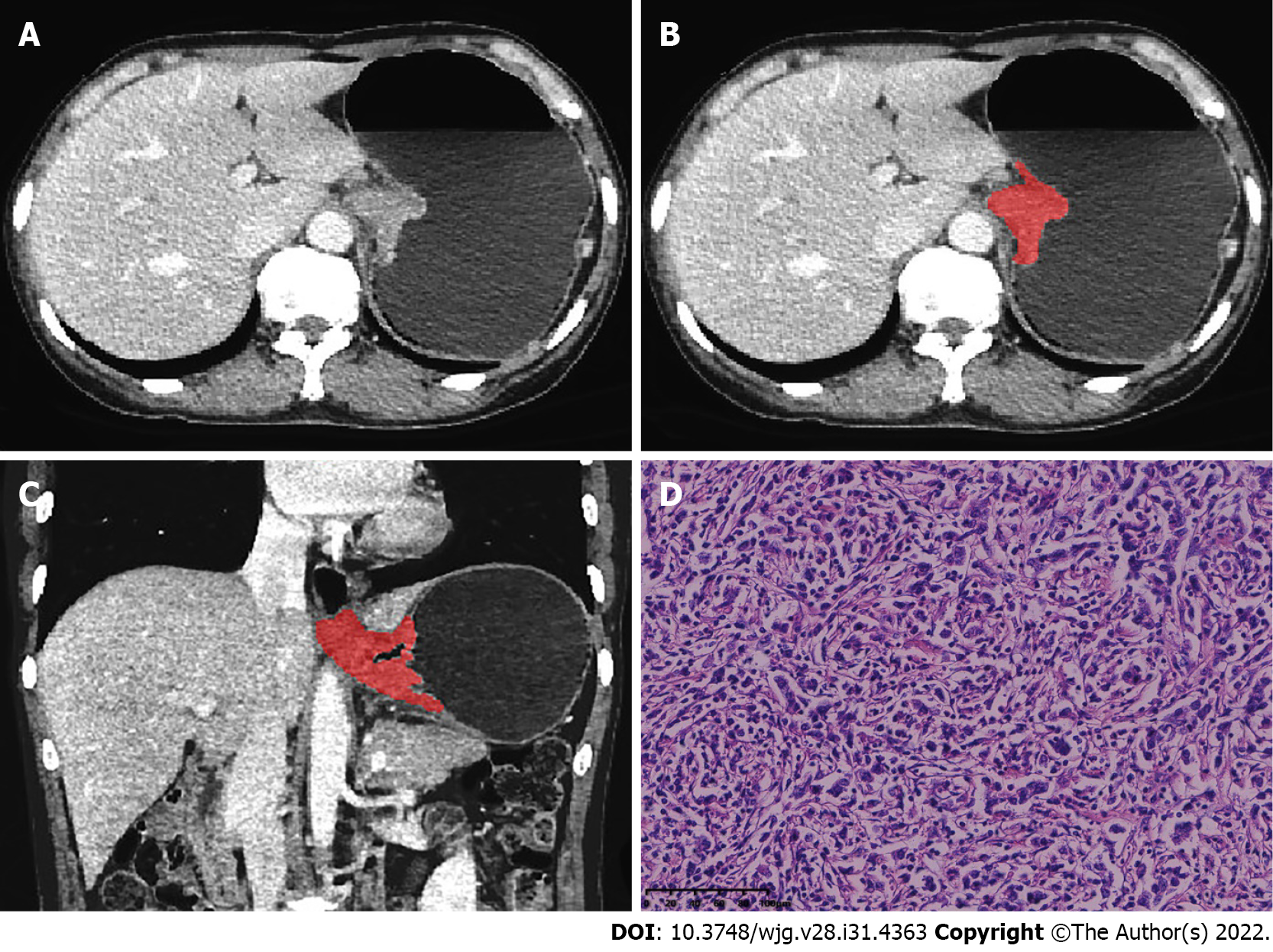
Figure 2 A 56-year-old man with adenocarcinoma of the esophagogastric junction.
A: Axial computed tomography image in venous phase; B: Schematic diagram of 2D region of interest (ROI) segmentation on ITK-SNAP software; C: Schematic diagram of 3D ROI segmentation on ITK-SNAP software; D: Postoperative pathological image confirming adenocarcinoma of the esophagogastric junction (HE staining, × 200).
- Citation: Du KP, Huang WP, Liu SY, Chen YJ, Li LM, Liu XN, Han YJ, Zhou Y, Liu CC, Gao JB. Application of computed tomography-based radiomics in differential diagnosis of adenocarcinoma and squamous cell carcinoma at the esophagogastric junction. World J Gastroenterol 2022; 28(31): 4363-4375
- URL: https://www.wjgnet.com/1007-9327/full/v28/i31/4363.htm
- DOI: https://dx.doi.org/10.3748/wjg.v28.i31.4363


