Copyright
©The Author(s) 2022.
World J Gastroenterol. Aug 14, 2022; 28(30): 4211-4220
Published online Aug 14, 2022. doi: 10.3748/wjg.v28.i30.4211
Published online Aug 14, 2022. doi: 10.3748/wjg.v28.i30.4211
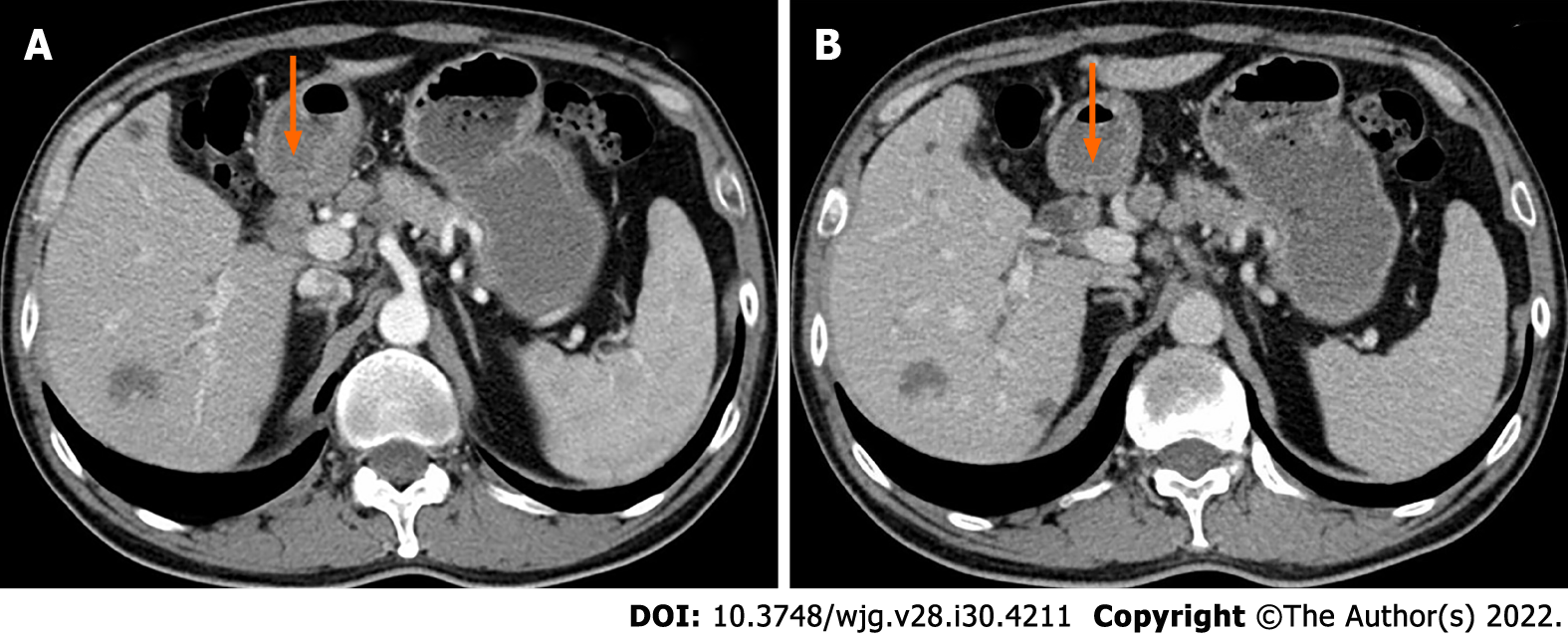
Figure 2 Contrast-enhanced computed tomography images of the patient.
Contrast-enhanced computed tomography showed a hypoenhancement nodule in the upper extrahepatic bile duct (orange arrow).
- Citation: Yuan ZQ, Yan HL, Li JW, Luo Y. Contrast-enhanced ultrasound of a traumatic neuroma of the extrahepatic bile duct: A case report and review of literature. World J Gastroenterol 2022; 28(30): 4211-4220
- URL: https://www.wjgnet.com/1007-9327/full/v28/i30/4211.htm
- DOI: https://dx.doi.org/10.3748/wjg.v28.i30.4211


