Copyright
©The Author(s) 2022.
World J Gastroenterol. Aug 14, 2022; 28(30): 4211-4220
Published online Aug 14, 2022. doi: 10.3748/wjg.v28.i30.4211
Published online Aug 14, 2022. doi: 10.3748/wjg.v28.i30.4211
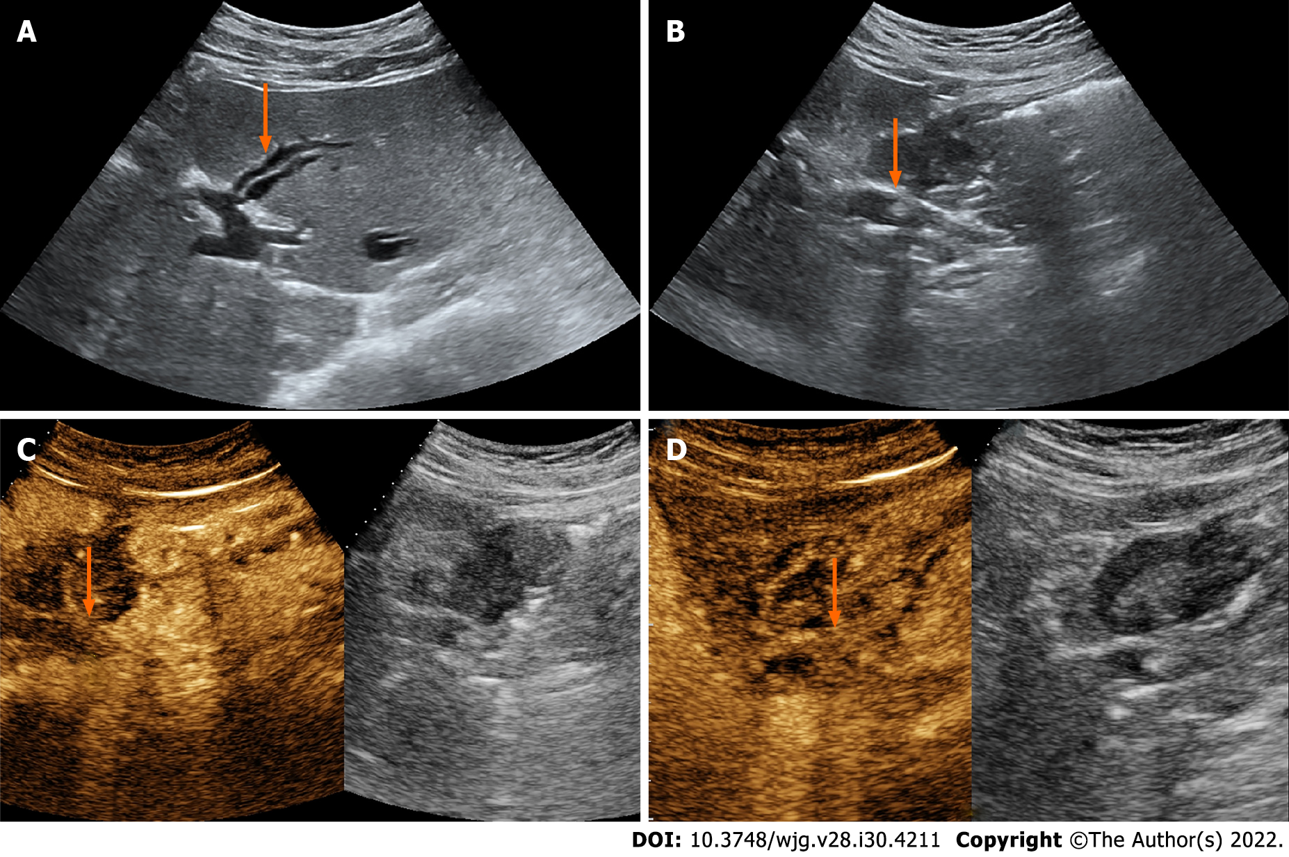
Figure 1 Ultrasound images of the patient.
A and B: The ultrasound (US) showed mild to moderate intrahepatic bile duct dilatation (orange arrow) and a hyperechoic nodule sized 0.8 cm × 0.6 cm (orange arrow) in the extrahepatic bile duct; C and D: In the arterial phase, contrast-enhanced US (CEUS) showed slight hyperenhancement (orange arrow); in the venous phase, CEUS showed isoenhancement (orange arrow).
- Citation: Yuan ZQ, Yan HL, Li JW, Luo Y. Contrast-enhanced ultrasound of a traumatic neuroma of the extrahepatic bile duct: A case report and review of literature. World J Gastroenterol 2022; 28(30): 4211-4220
- URL: https://www.wjgnet.com/1007-9327/full/v28/i30/4211.htm
- DOI: https://dx.doi.org/10.3748/wjg.v28.i30.4211


