Copyright
©The Author(s) 2022.
World J Gastroenterol. Aug 14, 2022; 28(30): 4044-4052
Published online Aug 14, 2022. doi: 10.3748/wjg.v28.i30.4044
Published online Aug 14, 2022. doi: 10.3748/wjg.v28.i30.4044
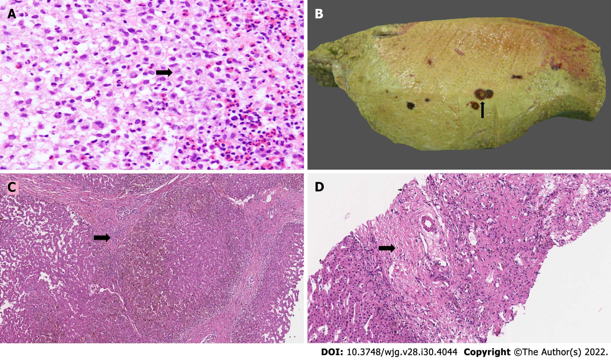
Figure 1 Langerhans cell histiocytosis exhibit diverse morphological features in the liver.
A: Aggregates of Langerhans cells admixed with inflammatory cells (arrow, H&E, 40 ×); B: Explant liver with dilated bile ducts and sludge (arrow); C: Liver wedge biopsy with evolving biliary cirrhosis and mild peripheral bile ductular proliferation (arrow, H&E, 20 ×); D: Liver biopsy with ductopenia (arrow, H&E, 40 ×).
- Citation: Menon J, Rammohan A, Vij M, Shanmugam N, Rela M. Current perspectives on the role of liver transplantation for Langerhans cell histiocytosis: A narrative review. World J Gastroenterol 2022; 28(30): 4044-4052
- URL: https://www.wjgnet.com/1007-9327/full/v28/i30/4044.htm
- DOI: https://dx.doi.org/10.3748/wjg.v28.i30.4044


