Copyright
©The Author(s) 2022.
World J Gastroenterol. Aug 7, 2022; 28(29): 3960-3970
Published online Aug 7, 2022. doi: 10.3748/wjg.v28.i29.3960
Published online Aug 7, 2022. doi: 10.3748/wjg.v28.i29.3960
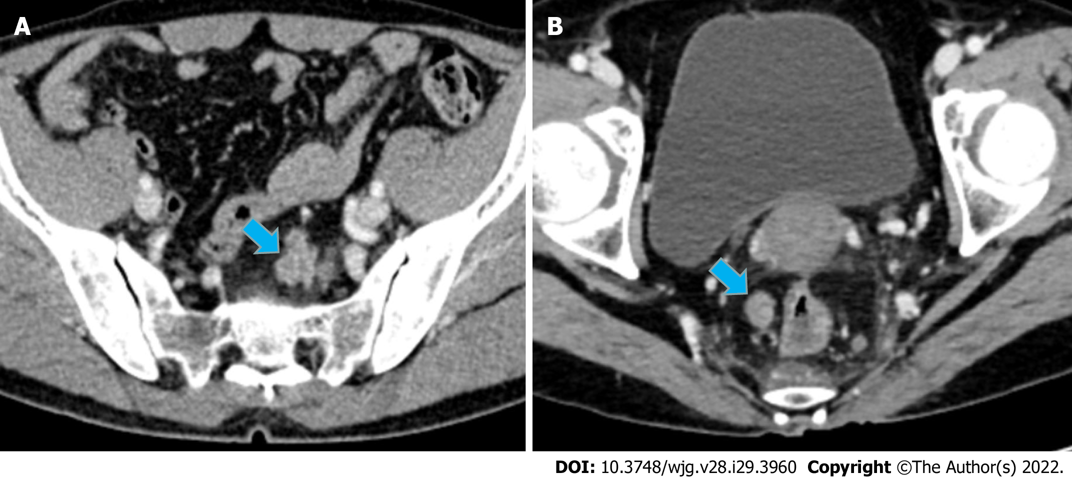
Figure 4 Case presentation.
A: A 56-year-old man with upper RC, the nodule of TDs (size: 24 mm × 16 mm) had an irregular shape; B: A 44-year-old man with lower RC, the nodule of TDs (size: 14 mm × 11 mm) had a regular oval shape. It is difficult to distinguish TDs and LNM from conventional imaging findings. For these two patients, Rad-score of the largest peritumoral nodule achieved correct diagnosis (values = 0.98 and 0.97, respectively). RC: Rectal cancer; TDs: Tumor deposits; LNM: Lymph node metastasis.
- Citation: Zhang YC, Li M, Jin YM, Xu JX, Huang CC, Song B. Radiomics for differentiating tumor deposits from lymph node metastasis in rectal cancer. World J Gastroenterol 2022; 28(29): 3960-3970
- URL: https://www.wjgnet.com/1007-9327/full/v28/i29/3960.htm
- DOI: https://dx.doi.org/10.3748/wjg.v28.i29.3960


