Copyright
©The Author(s) 2022.
World J Gastroenterol. Jul 28, 2022; 28(28): 3732-3738
Published online Jul 28, 2022. doi: 10.3748/wjg.v28.i28.3732
Published online Jul 28, 2022. doi: 10.3748/wjg.v28.i28.3732
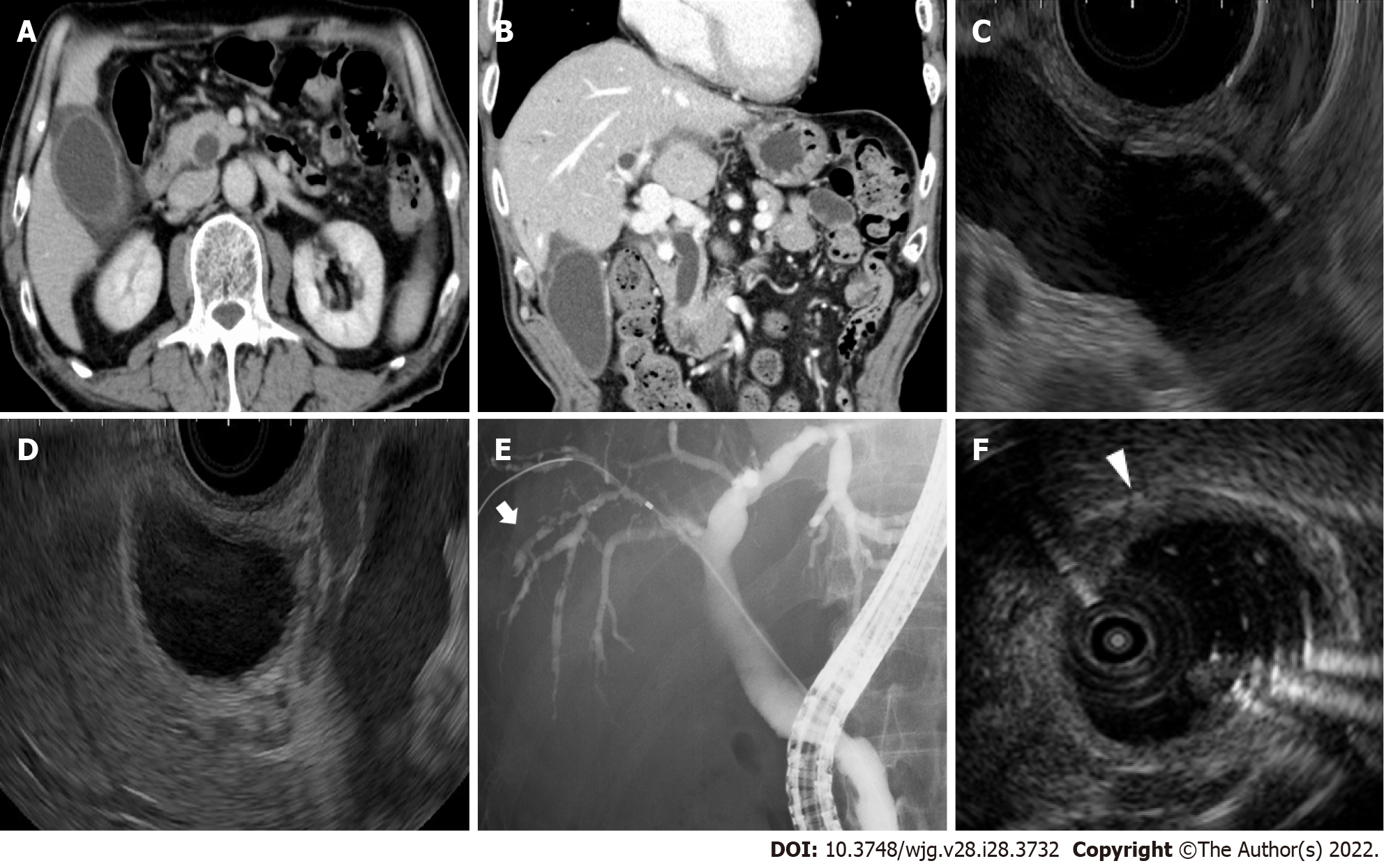
Figure 1 Imaging examinations of the gallbladder and common bile duct.
A, B: Contrast-enhanced computed tomography shows swelling and wall thickness of the gallbladder and common bile duct; C, D: Endoscopic ultrasonography shows dilatation of the common bile duct without obstruction and wall thickness of the gallbladder; E: Endoscopic retrograde cholangiopancreatography shows a dilated common bile duct and irregularly narrowed right intrahepatic bile duct (white arrow); F: Intraductal ultrasonography shows the wall thickness from the right bile duct to the common bile duct (white arrowhead).
- Citation: Tanaka T, Sakai A, Tsujimae M, Yamada Y, Kobayashi T, Masuda A, Kodama Y. Delayed immune-related sclerosing cholangitis after discontinuation of pembrolizumab: A case report. World J Gastroenterol 2022; 28(28): 3732-3738
- URL: https://www.wjgnet.com/1007-9327/full/v28/i28/3732.htm
- DOI: https://dx.doi.org/10.3748/wjg.v28.i28.3732


