Copyright
©The Author(s) 2022.
World J Gastroenterol. Jul 21, 2022; 28(27): 3488-3502
Published online Jul 21, 2022. doi: 10.3748/wjg.v28.i27.3488
Published online Jul 21, 2022. doi: 10.3748/wjg.v28.i27.3488
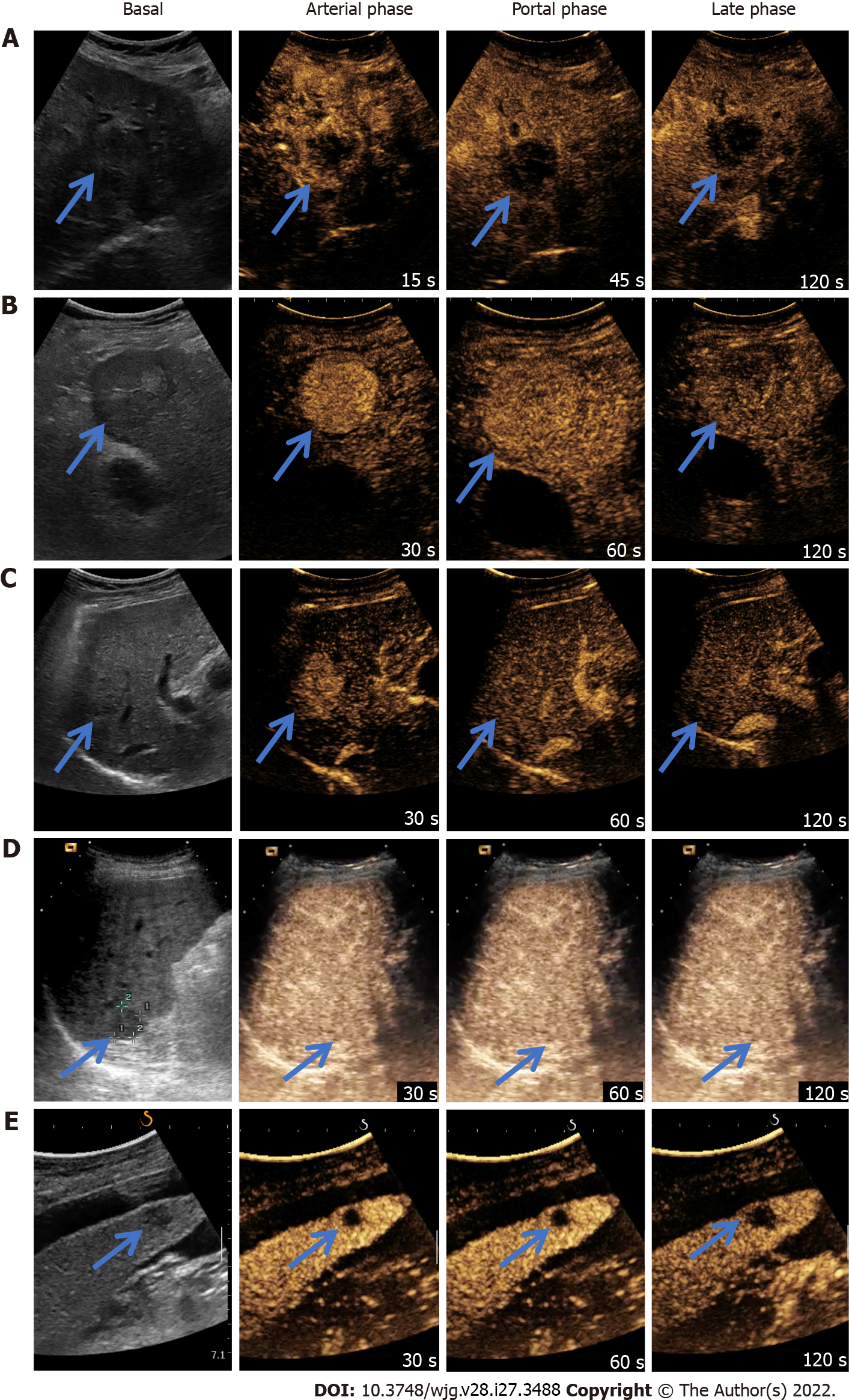
Figure 2 Examples of different contrast-enhanced ultrasound Liver Imaging Reporting and Data System classes.
A: Contrast-enhanced ultrasound (CEUS) LR-M. Notice rim arterial phase hyperenhancement and early washout, before 60 s; B: CEUS LR-5. Notice homogeneous arterial phase hyperenhancement, isoenhancement in portal phase, and mild washout in the late phase; C: CEUS LR-4. Notice homogeneous arterial phase hyperenhancement and isoenhancement in both portal and late phases; D: CEUS LR-3 iso-iso. Notice isoenhancement in all phases; E: CEUS LR-3 hypo-hypo. Notice hypoenhancement in all phases. The arrows show the target lesion.
- Citation: Vidili G, Arru M, Solinas G, Calvisi DF, Meloni P, Sauchella A, Turilli D, Fabio C, Cossu A, Madeddu G, Babudieri S, Zocco MA, Iannetti G, Di Lembo E, Delitala AP, Manetti R. Contrast-enhanced ultrasound Liver Imaging Reporting and Data System: Lights and shadows in hepatocellular carcinoma and cholangiocellular carcinoma diagnosis. World J Gastroenterol 2022; 28(27): 3488-3502
- URL: https://www.wjgnet.com/1007-9327/full/v28/i27/3488.htm
- DOI: https://dx.doi.org/10.3748/wjg.v28.i27.3488


