Copyright
©The Author(s) 2022.
World J Gastroenterol. Jul 21, 2022; 28(27): 3383-3397
Published online Jul 21, 2022. doi: 10.3748/wjg.v28.i27.3383
Published online Jul 21, 2022. doi: 10.3748/wjg.v28.i27.3383
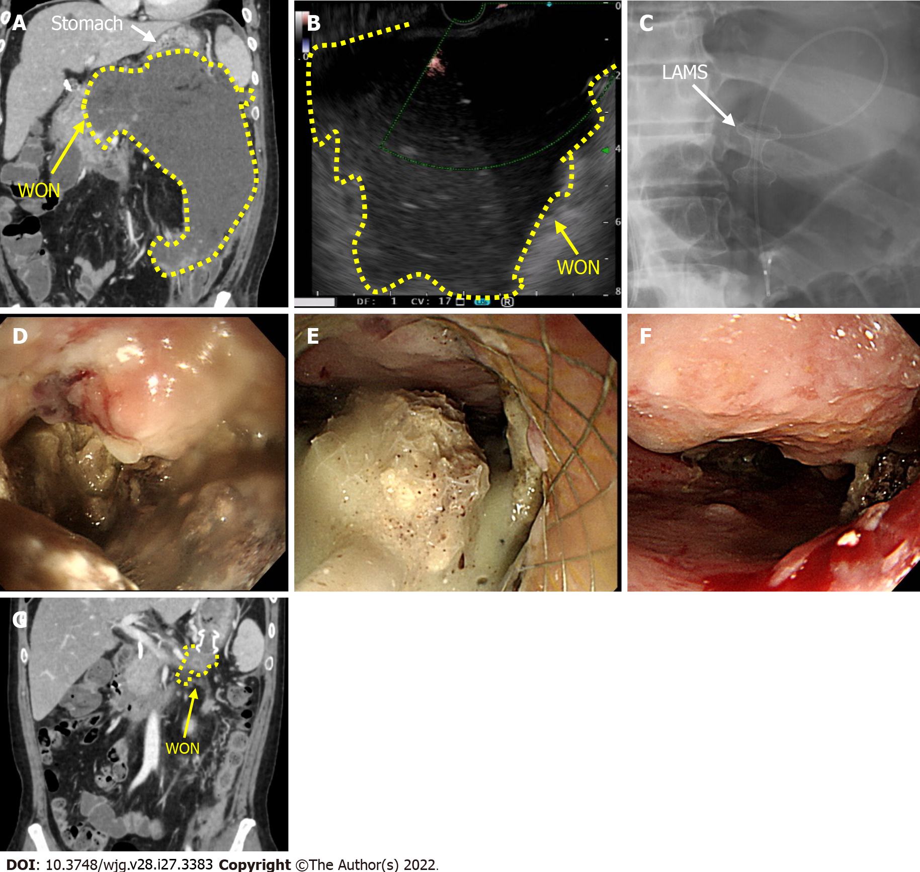
Figure 3 A case with endoscopic transluminal drainage with lumen-apposing metal stent.
A: Computed tomography (CT) scan before performing the endoscopic ultrasonography (EUS)-guided drainage (White arrow shows the stomach and the yellow arrow shows the walled-off necrosis (WON); the yellow dotted line is the demarcation line of the WON); B: EUS (with color doppler) picture shows marked echoic lesion without vessels; C: Lumen-apposing metal stent (LAMS) and nasobiliary drainage tube were placed (white arrow shows LAMS: Hot AXIOSTM 15 mm × 10 mm, Boston Scientific, Marlborough, MA, United States; Boston Scientific Japan, Tokyo, Japan); D: Esophagogastroduodenoscopy was inserted into necrotic cavity through LAMS; E: Necrosectomy was performed using endoscopic retrieval net; F: Endoscopic findings of the WON one month after the multiple necrosectomy sessions (2-3 times/wk); G: CT scan shows marked reduction of WON cavity one month after multiple necrosectomy sessions. WON: Walled-off necrosis; LAMS: Lumen-apposing metal stent.
- Citation: Purschke B, Bolm L, Meyer MN, Sato H. Interventional strategies in infected necrotizing pancreatitis: Indications, timing, and outcomes. World J Gastroenterol 2022; 28(27): 3383-3397
- URL: https://www.wjgnet.com/1007-9327/full/v28/i27/3383.htm
- DOI: https://dx.doi.org/10.3748/wjg.v28.i27.3383


