Copyright
©The Author(s) 2022.
World J Gastroenterol. Jul 14, 2022; 28(26): 3177-3200
Published online Jul 14, 2022. doi: 10.3748/wjg.v28.i26.3177
Published online Jul 14, 2022. doi: 10.3748/wjg.v28.i26.3177
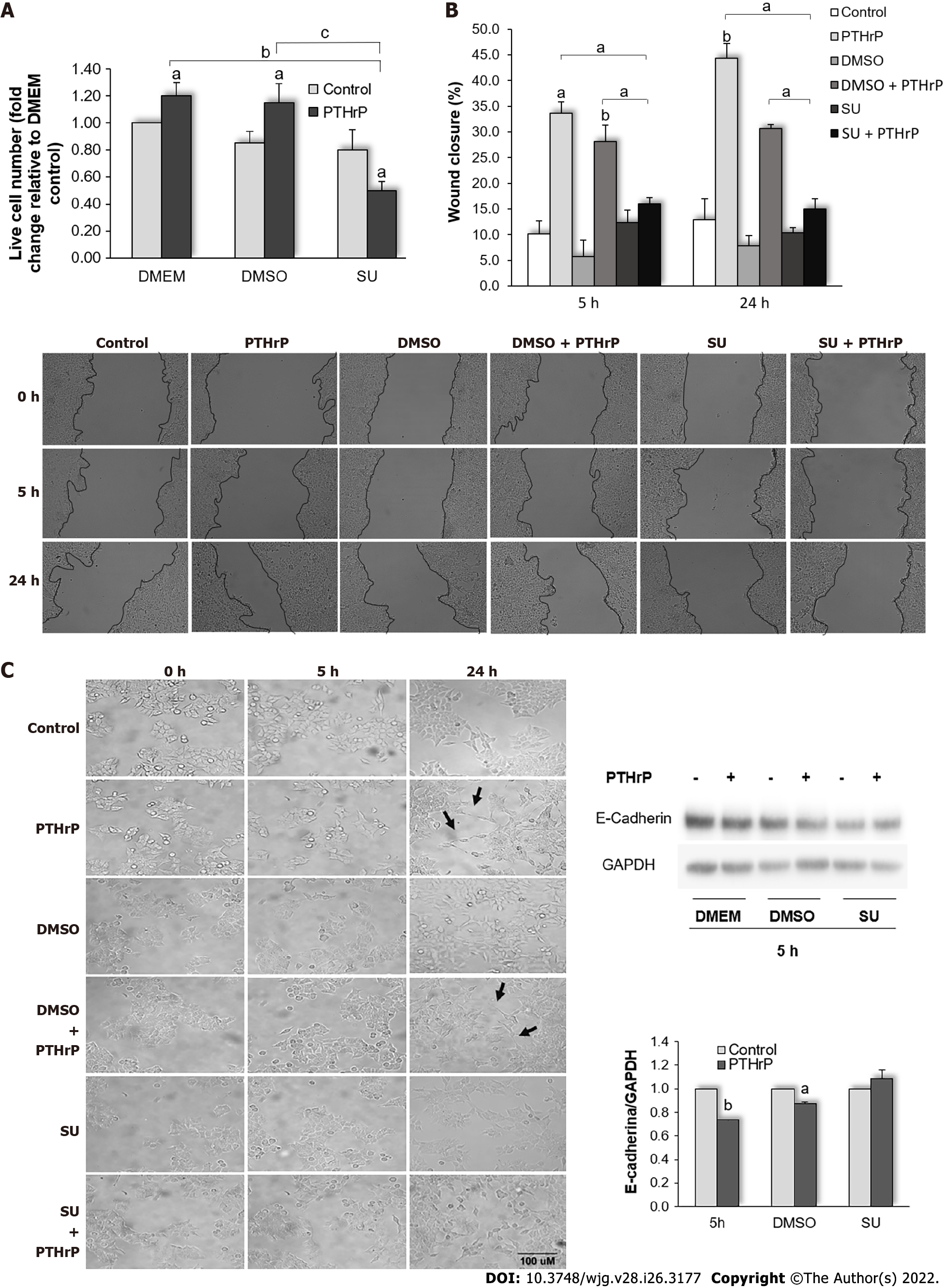
Figure 5 Parathyroid hormone-related peptide promotes events related to the aggressive behavior of HCT116 cells through the Met signaling pathway.
HCT116 cells were pre-incubated with SU11274, a specific Met inhibitor, for 30 min and then treated with or without parathyroid hormone-related peptide (PTHrP; 10-8M). A: Trypan blue technique showed that Met inhibition decreased the cell proliferation induced by PTHrP at 24 h; B: Images from wound healing assay show that Met inhibition reverted the wound closure promoted by PTHrP at 5 h and 24 h; C: E-cadherin protein levels analyzed by Western blot to investigate whether Met is involved in the decrease of this epithelial-mesenchymal transition (EMT) program marker induced by PTHrP in HCT116 cells. Using the Image J-NIH program, we performed the analysis of the parameters related to cell morphology. The arrows indicate the morphological changes corresponding to the transition from a polygonal structure to spindle-like structure related to EMT program progress observed when the cells were treated for 24 h with PTHrP or PTHrP plus dimethylsulfoxide (DMSO). In all experiments, a control with DMSO, the vehicle of the inhibitor, was performed. Graph bars represent the average of the results obtained from two independent experiments. DMEM: Dulbecco’s Modified Eagle Culture Medium; DMSO: Dimethylsulfoxide; EMT: Epithelial to mesenchymal transition; PTHrP: Parathyroid hormone-related peptide. aP < 0.05; bP < 0.01; cP < 0.001.
- Citation: Novoa Díaz MB, Carriere P, Gigola G, Zwenger AO, Calvo N, Gentili C. Involvement of Met receptor pathway in aggressive behavior of colorectal cancer cells induced by parathyroid hormone-related peptide. World J Gastroenterol 2022; 28(26): 3177-3200
- URL: https://www.wjgnet.com/1007-9327/full/v28/i26/3177.htm
- DOI: https://dx.doi.org/10.3748/wjg.v28.i26.3177


