Copyright
©The Author(s) 2022.
World J Gastroenterol. Jul 14, 2022; 28(26): 3008-3026
Published online Jul 14, 2022. doi: 10.3748/wjg.v28.i26.3008
Published online Jul 14, 2022. doi: 10.3748/wjg.v28.i26.3008
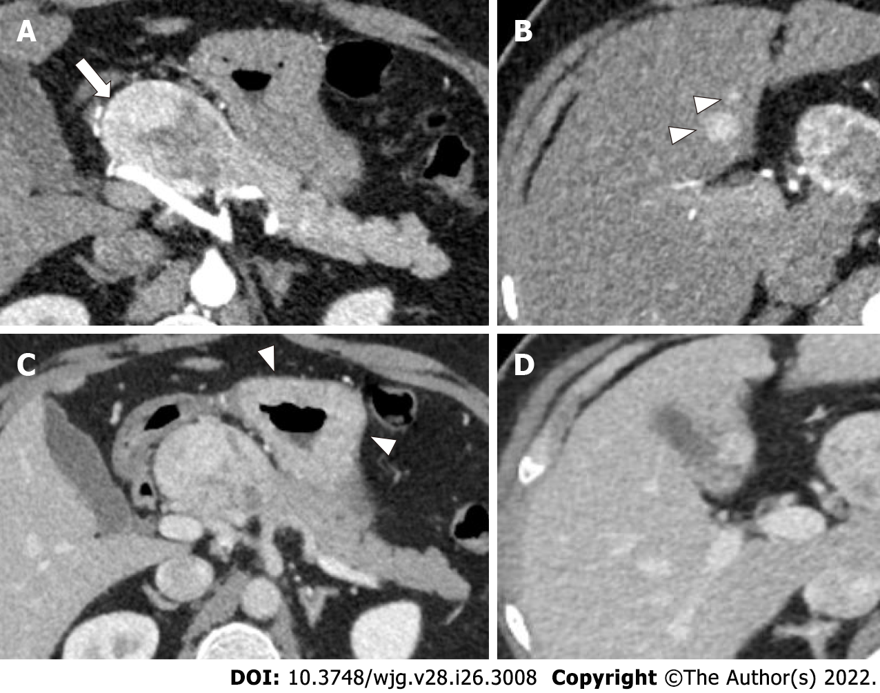
Figure 1 49-year-old man with epigastric pain and raised serum gastrin levels.
A and B: Axial contrast enhanced pancreatic phase computed tomography (CT) images show a well-defined hyperenhancing mass (arrow in A) in the head and neck of pancreas, abutting the proper hepatic artery along with two hyperenhancing focal lesions in the liver (arrowheads in B), indicating hepatic metastases; C and D: Axial portal venous phase CT images show retention of contrast in the lesions in both locations. Thickened gastric mucosal folds is also noted (arrowheads in C).
- Citation: Ramachandran A, Madhusudhan KS. Advances in the imaging of gastroenteropancreatic neuroendocrine neoplasms. World J Gastroenterol 2022; 28(26): 3008-3026
- URL: https://www.wjgnet.com/1007-9327/full/v28/i26/3008.htm
- DOI: https://dx.doi.org/10.3748/wjg.v28.i26.3008


