Copyright
©The Author(s) 2022.
World J Gastroenterol. Jul 7, 2022; 28(25): 2994-3000
Published online Jul 7, 2022. doi: 10.3748/wjg.v28.i25.2994
Published online Jul 7, 2022. doi: 10.3748/wjg.v28.i25.2994
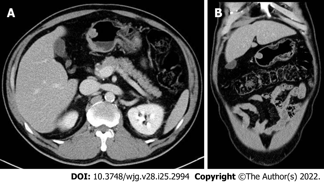
Figure 3 Abdominal computed tomography images.
Initial axial (A) and coronal (B) computed tomography images showing a 15 mm protruding intraluminal mass at gastric antrum.
- Citation: Cho JH, Lee SH. Early gastric cancer presenting as a typical submucosal tumor cured by endoscopic submucosal dissection: A case report. World J Gastroenterol 2022; 28(25): 2994-3000
- URL: https://www.wjgnet.com/1007-9327/full/v28/i25/2994.htm
- DOI: https://dx.doi.org/10.3748/wjg.v28.i25.2994


