Copyright
©The Author(s) 2022.
World J Gastroenterol. Jun 28, 2022; 28(24): 2748-2757
Published online Jun 28, 2022. doi: 10.3748/wjg.v28.i24.2748
Published online Jun 28, 2022. doi: 10.3748/wjg.v28.i24.2748
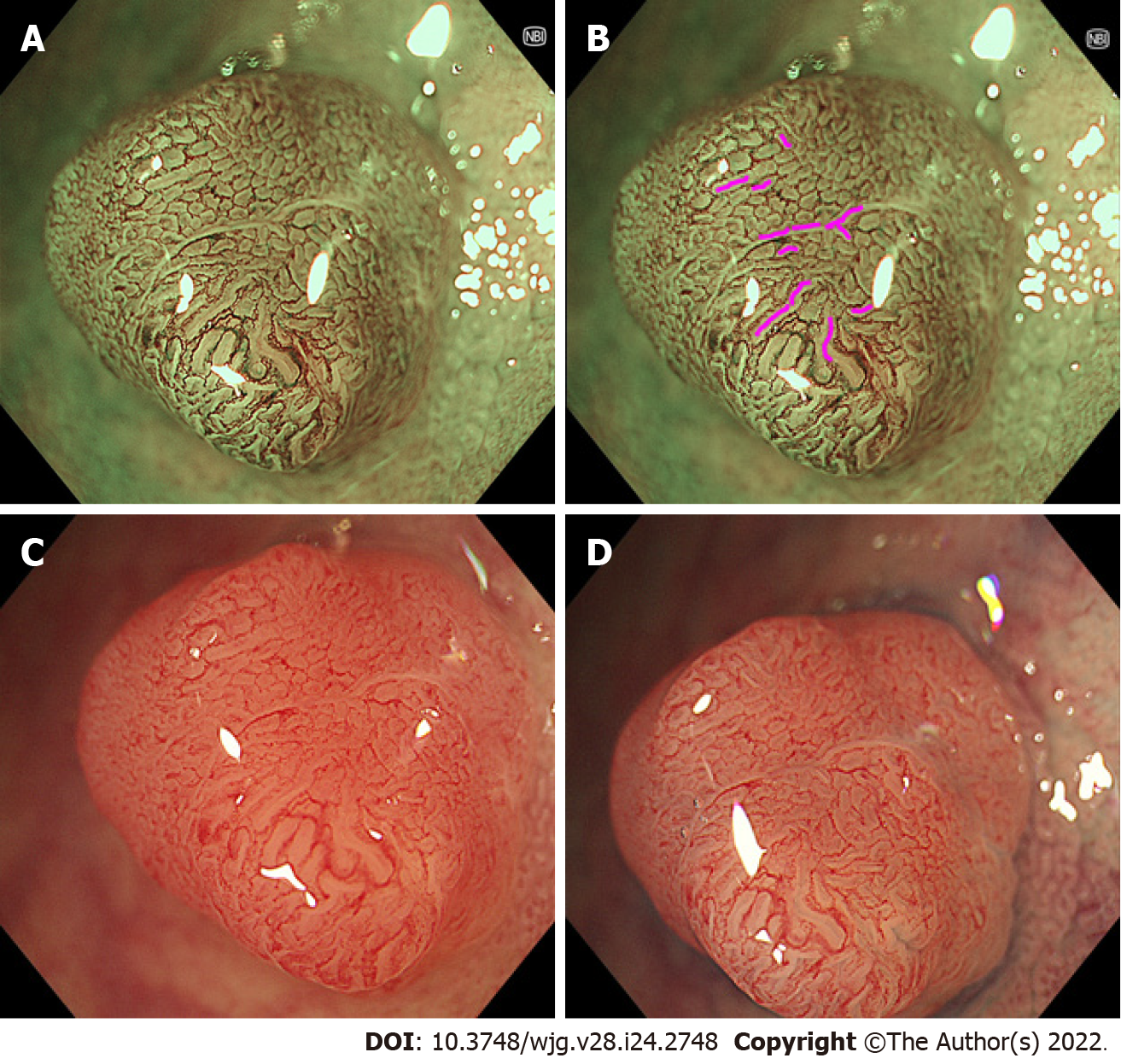
Figure 1 Representative images of “brown slits” in a low-grade tubular adenoma using conventional magnifying endoscopy.
A set of a conventional magnifying endoscope (CF-HQ290Z), X1 system, and 4K 32-inch monitor was used. A, B: Full-zoom (95 ×) magnification, narrow band imaging. Brown slits were observed inside and along the tubular glands surrounded by the microvessels; C: Full-zoom magnifying, white-light imaging observation without indigo carmine spraying showed faint and pale red slits inside and along the tubular glands; D: Full-zoom magnifying, white light imaging with indigo carmine spraying showed indigo carmine accumulated in the site corresponding to the brown slits.
- Citation: Toyoshima O, Nishizawa T, Yoshida S, Watanabe H, Odawara N, Sakitani K, Arano T, Takiyama H, Kobayashi H, Kogure H, Fujishiro M. Brown slits for colorectal adenoma crypts on conventional magnifying endoscopy with narrow band imaging using the X1 system. World J Gastroenterol 2022; 28(24): 2748-2757
- URL: https://www.wjgnet.com/1007-9327/full/v28/i24/2748.htm
- DOI: https://dx.doi.org/10.3748/wjg.v28.i24.2748


