Copyright
©The Author(s) 2022.
World J Gastroenterol. Jun 21, 2022; 28(23): 2609-2624
Published online Jun 21, 2022. doi: 10.3748/wjg.v28.i23.2609
Published online Jun 21, 2022. doi: 10.3748/wjg.v28.i23.2609
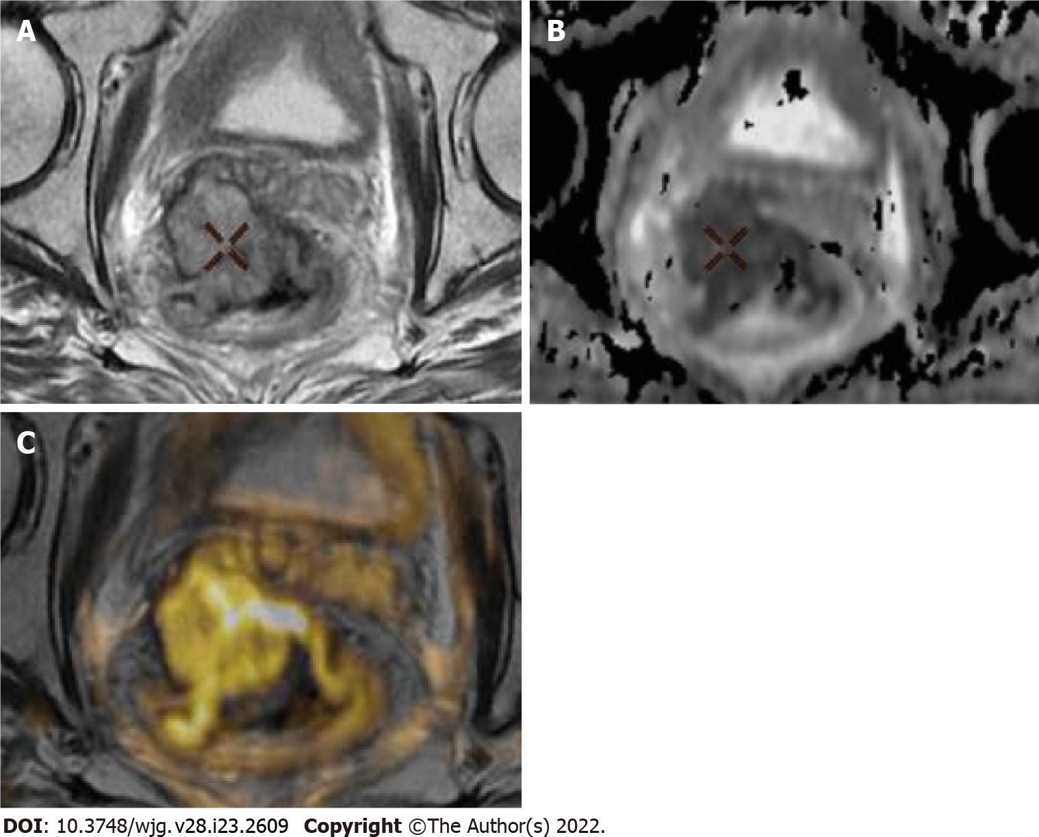
Figure 1 Representative images of magnetic resonance imaging protocol.
A-C: Axial T2 (A), apparent diffusion coefficient (ADC) map and T2 fusion ADC map color (B) and images of bulky tumor (C), showing tumor extending more than 5 mm into the mesorectal fat and invading the mesorectal fascia.
- Citation: Jiménez de los Santos ME, Reyes-Pérez JA, Domínguez Osorio V, Villaseñor-Navarro Y, Moreno-Astudillo L, Vela-Sarmiento I, Sollozo-Dupont I. Whole lesion histogram analysis of apparent diffusion coefficient predicts therapy response in locally advanced rectal cancer. World J Gastroenterol 2022; 28(23): 2609-2624
- URL: https://www.wjgnet.com/1007-9327/full/v28/i23/2609.htm
- DOI: https://dx.doi.org/10.3748/wjg.v28.i23.2609


